Vertebral compression injuries comprise nonstructural fractures (A0), wedge-compression fractures (A1), split fractures (A2), incomplete burst fractures (A3) and complete burst fractures (A4). In acute trauma, they result from axial loading forces, but can develop insidiously in vertebrae weakened by osteoporosis, metastatic disease, or infection. Alcohol, steroids, cytotoxic drugs, thyroid hormones, and heparin all predispose to osteoporosis and compression fractures. An isolated compression injury increases the risk of subsequent fractures due to altered biomechanics of the kyphotic spine.
On the normal AP radiograph, the pedicles should be intact, and the interpedicular and interspinous distances should gradually increase from craniad to caudad. Focal widening of the interpedicular distance indicates a compression-burst type fracture whereas focal increase in the interspinous distance suggests a distraction injury. These findings should prompt CT, and if there are associated neurologic deficits, MRI of the spine.
Nonstructural fractures are minor injuries that include spinous and transverse process fractures and do not compromise the stability of the spine.
Wedge-compression fractures involve an endplate of the vertebral body but do not extend to the posterior wall. They appear on lateral radiographs as vertebral body wedge deformities. Height loss can be documented as the ratio of the ventral height of the involved vertebral body compared to the average of the heights of the two adjacent vertebrae. Treatment is usually conservative ( mobilization, analgesia, physiotherapy), but operative treatment may be needed in cases with > 15% angulation.
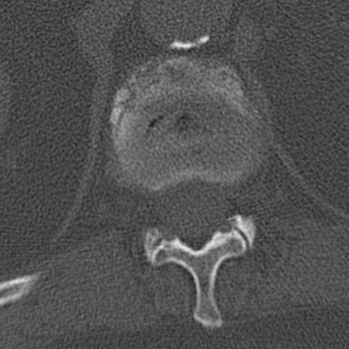
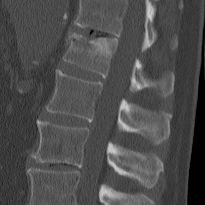
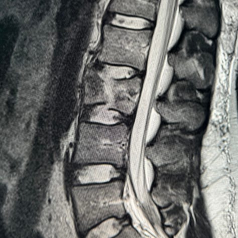
T12 wedge-compression fracture. Anterior superior compression, sclerosis, and fragmentation with ~ 20% loss of anterior vertebral body height. No distortion of the posterior vertebral body wall.
Split fractures are coronal fractures through both endplates without extension to the posterior vertebral body wall. Most are treated conservatively unless there is wide separation of the fragments.
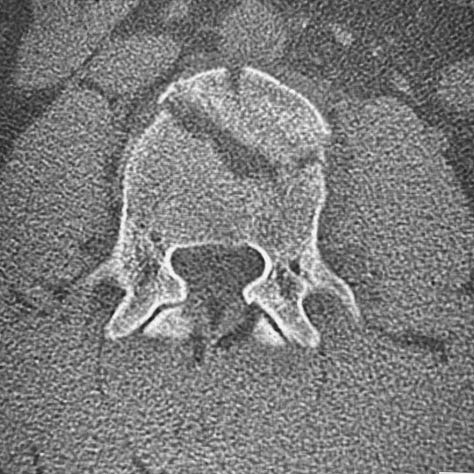
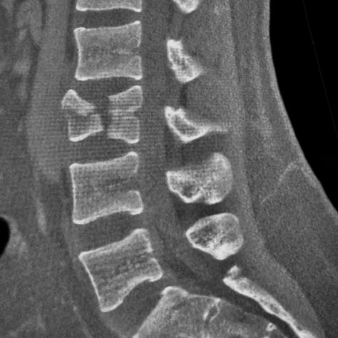
Pincer type (split) compression fracture. Coronal split with sparing of the posterior vertebral cortex.
Incomplete and complete burst fractures always involve the posterior vertebral body wall, which normally forms a smooth arch defining the anterior aspect of the spinal canal. In burst fractures, it is flattened or disrupted, with retropulsed fragments that can narrow the canal. Posterior element (pedicle, lamina, and spinous process) fractures are often present in more severe injuries. Burst fractures can occur at any spinal level, but are most frequently seen in the cervical spine and near the thoracolumbar junction. An incomplete burst fracture (A3) involves only the superior endplate, whereas a complete burst fracture (A4) involves both endplates.
Axial CT images typically show a “double cortex,” paraspinous hematoma, distortion of the posterior cortex (in burst fractures) and, if present, laminar and pedicle fractures. Sagittal reformations best demonstrate the extent of middle column retropulsion, vertebral body height loss and spinal canal narrowing. Coronal reformations show lateral height loss and angulation. When reporting a compression fracture, one should include an estimate of angulation, height loss, and any spinal canal compromise. MRI should be performed in any patient with neurologic compromise.
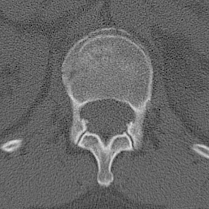
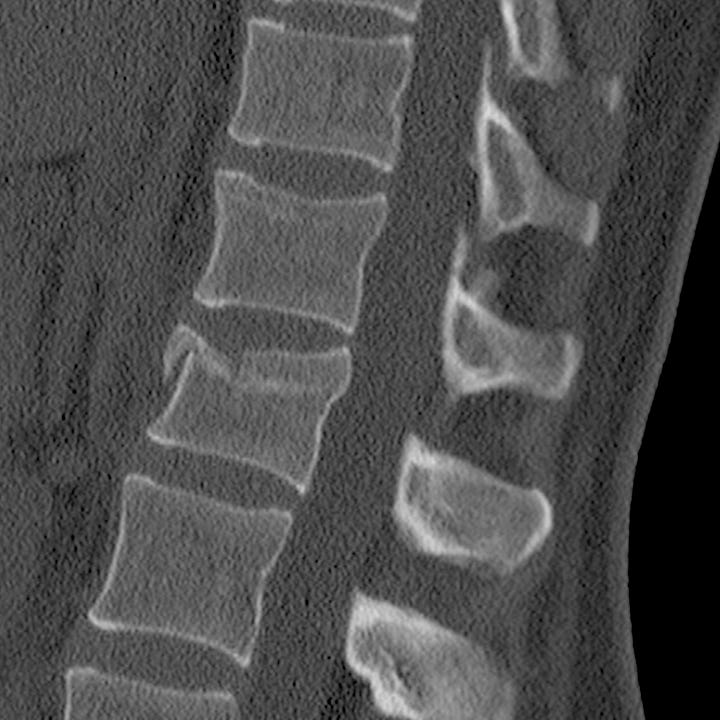
L1 incomplete burst fracture. Anterior compression with “double cortex” sign. Flattening of the anteri- or spinal canal wall and slight posterior bulge of the upper vertebral body indicate middle vertebral column involvement and potential instability. The inferior cortex is intact.
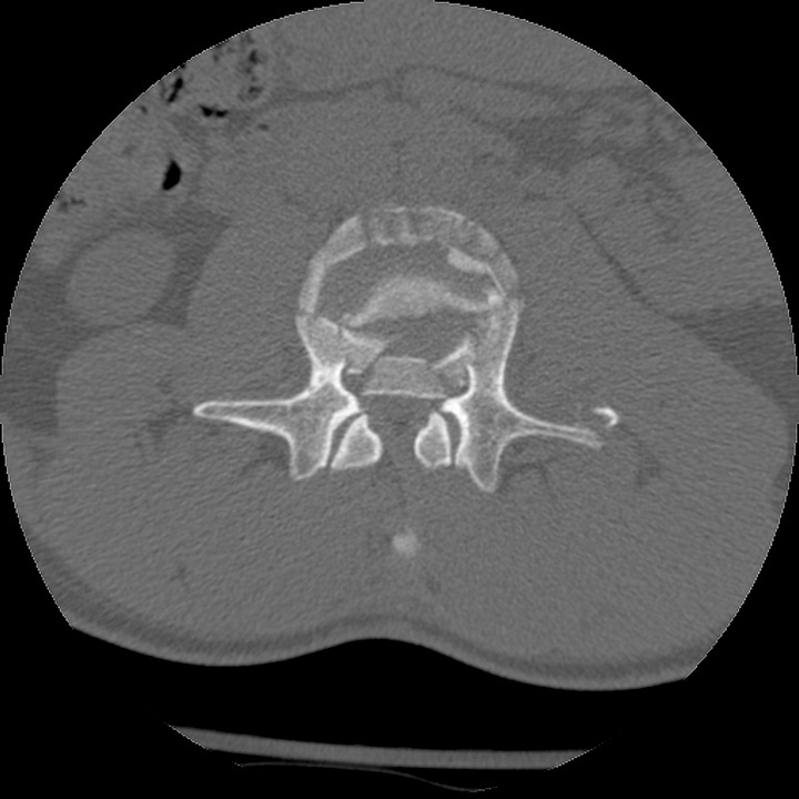
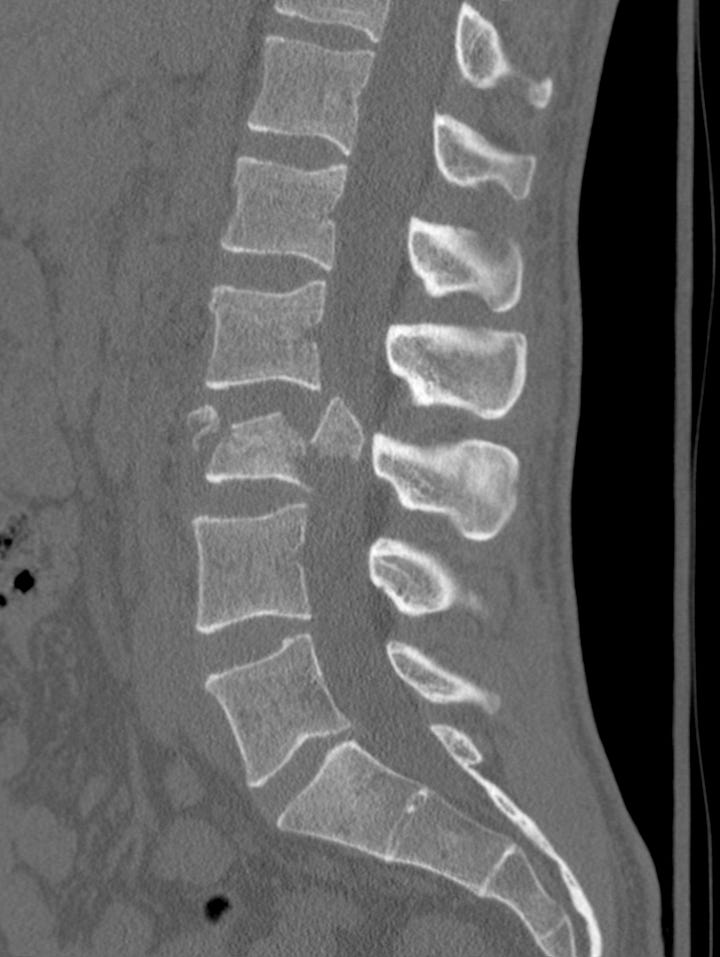
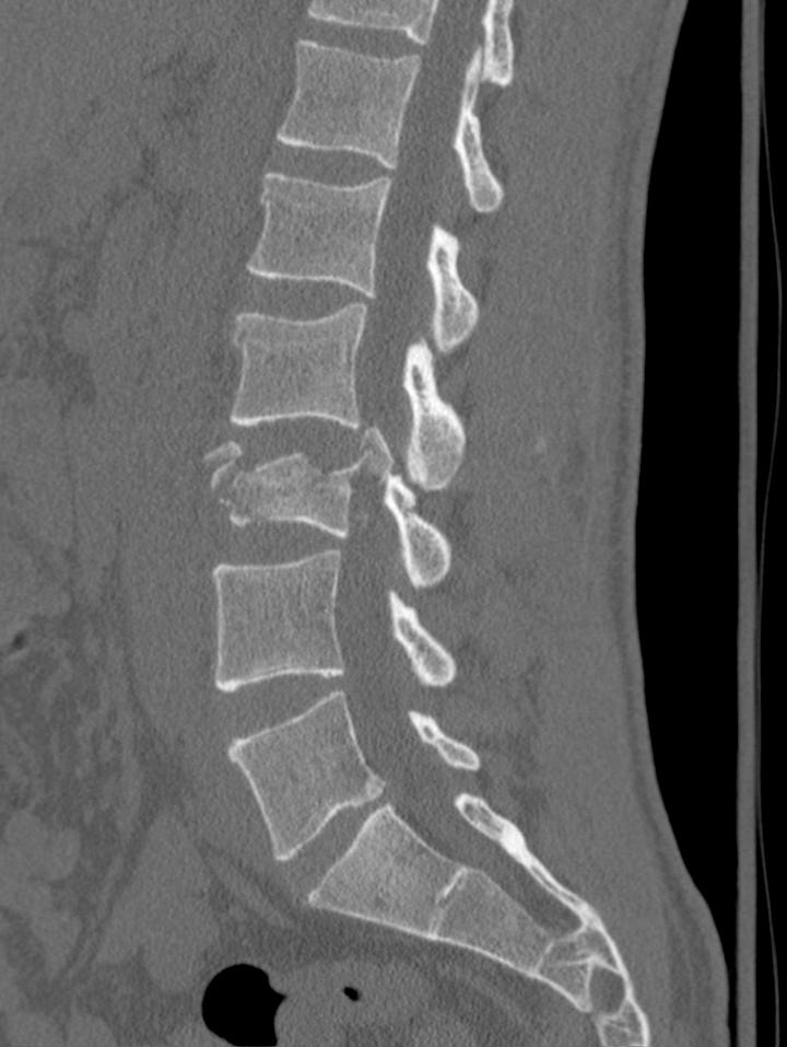
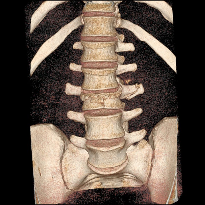
L3 complete burst fracture. Unstable, highly comminuted fracture involving both superior and inferior vertebral cortices, large retropulsed fragment, left transverse process fracture. ~ 50% height loss.

