Traumatic subarachnoid hemorrhage results either from direct pial vascular injury or extension from a hemorrhagic cortical contusion, parenchymal hematoma, or extra-axial hematoma. Although it is often seen in association with other brain injuries, it can be subtle, particularly if it is the only imaging finding in a patient with known head trauma. In contrast to subdural or epidural hematomas, blood that collects between the pia and the arachnoid membrane fills cerebral sulci and appears as fine linear or serpentine juxtacortical densities. Hemorrhage is also commonly seen in the perimesencephalic cisterns, particularly the interpeduncular and ambient cisterns. Over a period of days, blood becomes isodense to brain and ultimately resolves.
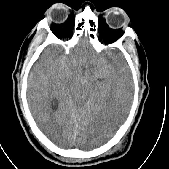

Isolated traumatic subarachnoid hemorrhage. Cerebral swelling with effacement of perimesencephalic cisterns and cortical sulci. Subarachnoid hemorrhage is present in the interpeduncular and ambient cisterns as well as several frontal and parietal convexity sulci.
It is important to keep in mind that the accident leading to a patient’s head injury may have been precipitated by loss of consciousness due to a ruptured aneurysm, arteriovenous malformation, or other non-traumatic condition. Blood that fills the suprasellar and middle cerebral artery cisterns or extends to the lateral (Sylvian) fissures in the absence of other traumatic findings (contusion, subdural hematoma, epidural hematoma), is most likely due to aneurysmal or other primary hemorrhage. In this case, the intracranal vasculature should be further evaluated by CT or conventional angiography.
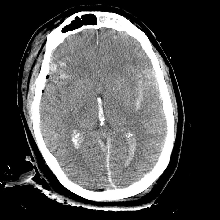
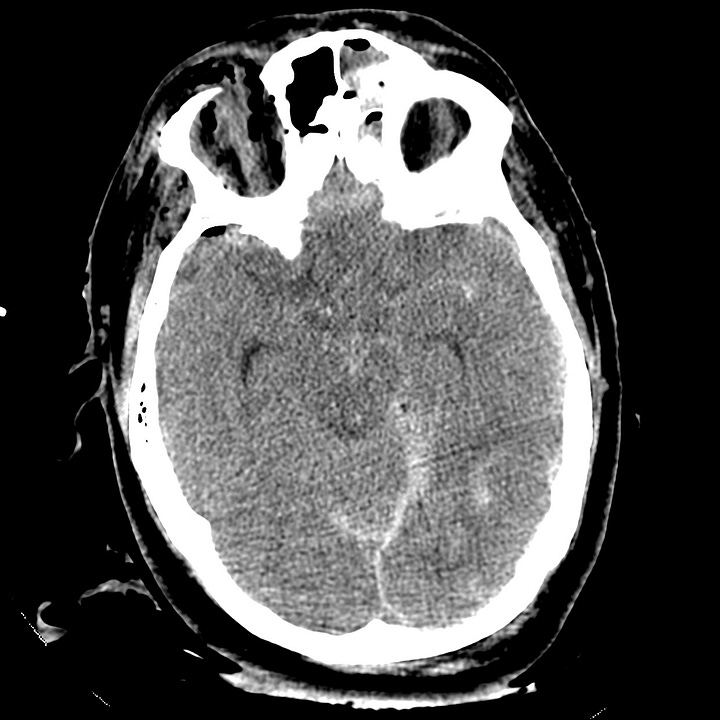
Traumatic subarachnoid hemorrhage associated with other cerebral injuries. Generalized cerebral swelling with right frontal cortical contusions, pneumocephalus, clot within the third and lateral ventricles, and small falcine/tentorial subdural hematoma. Traumatic subarachnoid hemorrhage is present in the interpeduncular cistern and left Sylvian fissure.
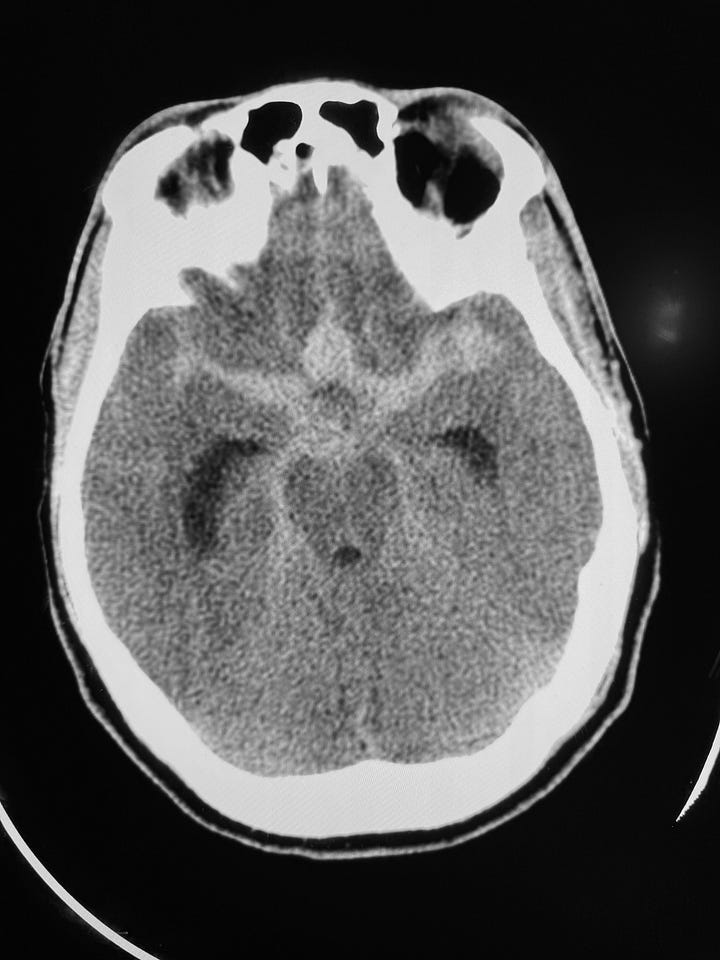
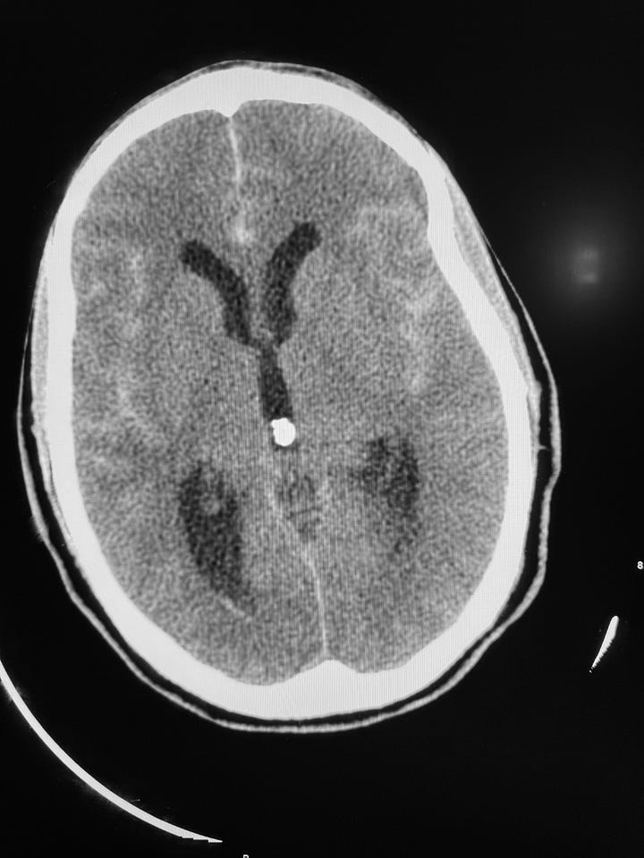
Subarachnoid hemorrhage due to ruptured anterior communicating artery (for comparison). Large amount of blood in suprasellar, perimesencephalic, and middle cerebral artery cisterns. Hemorrhage is also present in the anterior inter-hemispheric fissure and Sylvian fissures. Hydrocephalus with enlarged third and lateral ventricles. Small amount of intraventricular hemorrhage. This pattern should not be considered traumatic.
Subarachnoid hemorrhage is associated with increased morbidity and mortality in trauma, reflecting its potential to induce vasospasm and its frequent association with more severe mechanisms of head injury. Subarachnoid blood is also toxic to the adjacent cortex and can interfere with normal CSF absorption, which can lead to late complications of chronic communicating hydrocephalus or superficial siderosis.

