Blunt or penetrating chest trauma can result in aortic injury that ranges from minimal intimal damage to complete transection and exsanguination. Causes include high-speed motor vehicle accidents, falls, and penetrating trauma. In most blunt impact mechanisms, the comparatively mobile heart and ascending aorta accelerate relative to the descending aorta, which is fixed to the spine. The aorta stretches or tears at a point just distal to the origin of the left subclavian artery. Other potential sites of injury are the proximal descending aorta, aortic root, and distal descending aorta.
Patients with laceration of all three layers of the aortic wall invariably exsanguinate before reaching a hospital. If a patient with traumatic aortic injury survives to reach the emergency room, it is because the tear is transverse rather than longitudinal and the outer layer of the aorta (adventitia) remains intact.
Signs of a juxta-aortic hematoma on supine portable radiographs include mediastinal widening, indistinct margins, poor aortic contour definition, a widened paratracheal stripe, pleural effusion, caudal displacement of the right mainstem bronchus, and rightward displacement of an oro- or nasogastric tube. Many patients will have no mediastinal abnormality on portable chest radiograph.
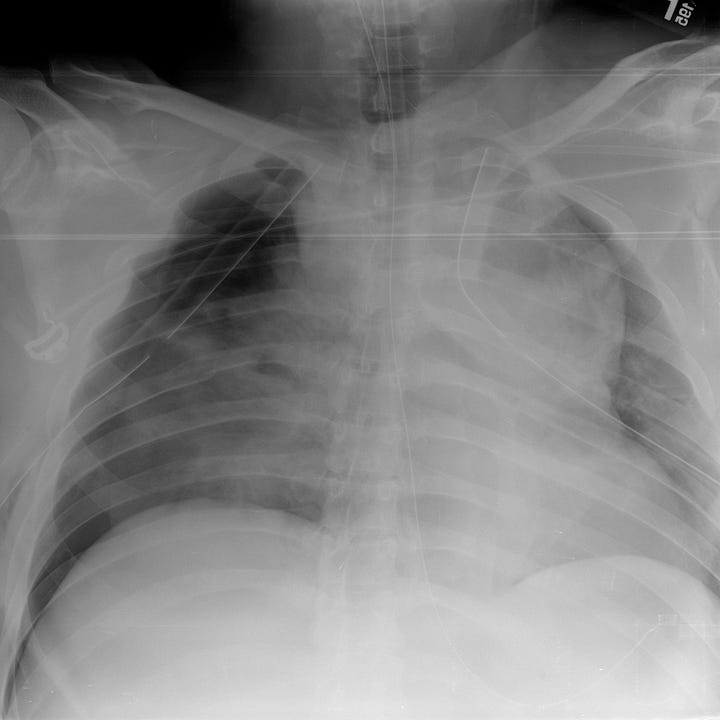
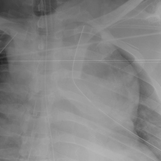
Traumatic aortic injury (11-story fall). Marked upper mediastinal widening with sharply-defined lateral border. Bilateral airspace opacities indicate pulmonary contusion or aspiration. Left scapular body fracture.
CT in arterial phase should be performed in patients with reported high-energy injury mechanism or who have upper rib, sternal, or scapular fractures on portable radiograph. A normal thoracic CT reliably excludes great vessel injury and can differentiate true aortic disruption from venous mediastinal hematoma by identifying direct findings of aortic injury: pseudoaneurysm, intimal flap, mural thrombus, arterial contrast extravasation, and focal change in aortic contour (“pseudocoarctation”).
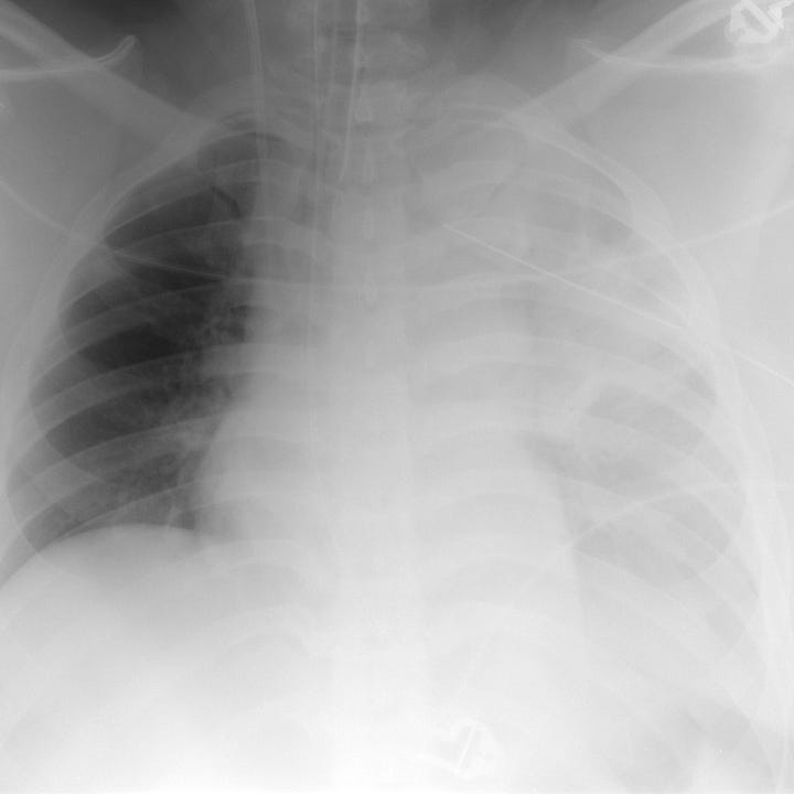
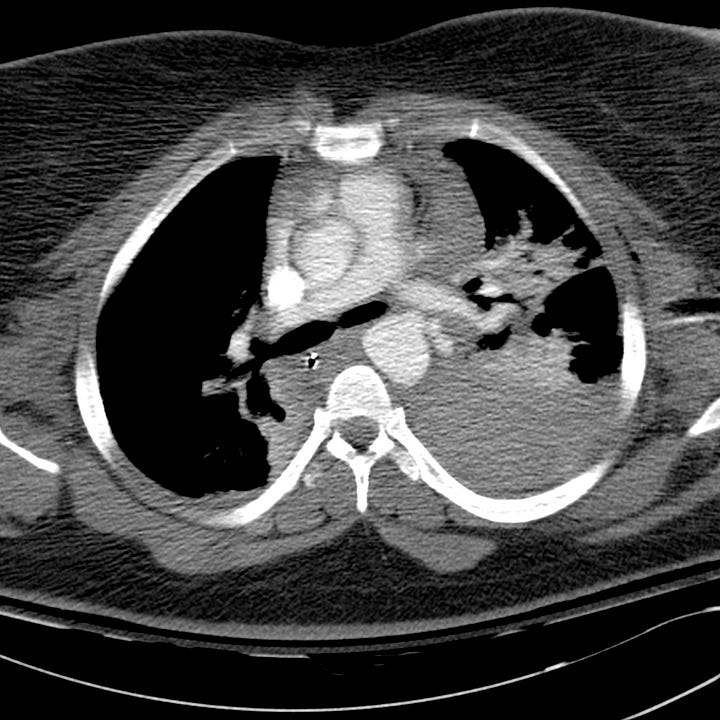
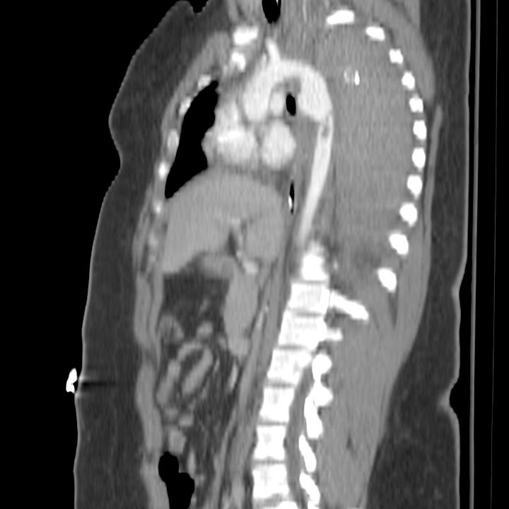

Traumatic aortic injury with large hemothorax. Portable supine radiograph shows widened, ill-defined upper mediastinum. The left hemithorax is opacified. The orogastric tube is displaced to the right. Postcontrast CT demonstrates a large left hemothorax and hemomediastinum. A small aortic pseudoaneurysm is visible adjacent to the left pulmonary artery on the axial image. Sagittal reformations show an associated aortic intramural hematoma, intimal flap, and pseudocoarctation.


