Shock bowel describes the intestinal CT findings seen in trauma patients who are initially hypotensive and hypovolemic; diffuse small-bowel wall thickening, fluid-filled, dilated small-bowel loops, and pronounced mucosal enhancement. The large bowel usually appears normal. Other CT findings related to systemic hypotension or hypovolemia include a flattened inferior vena cava (IVC), small-caliber aorta, and intense renal enhancement, the latter due to hypoperfusion. These generally resolve completely with effective fluid management.
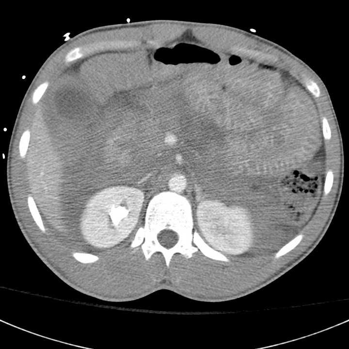
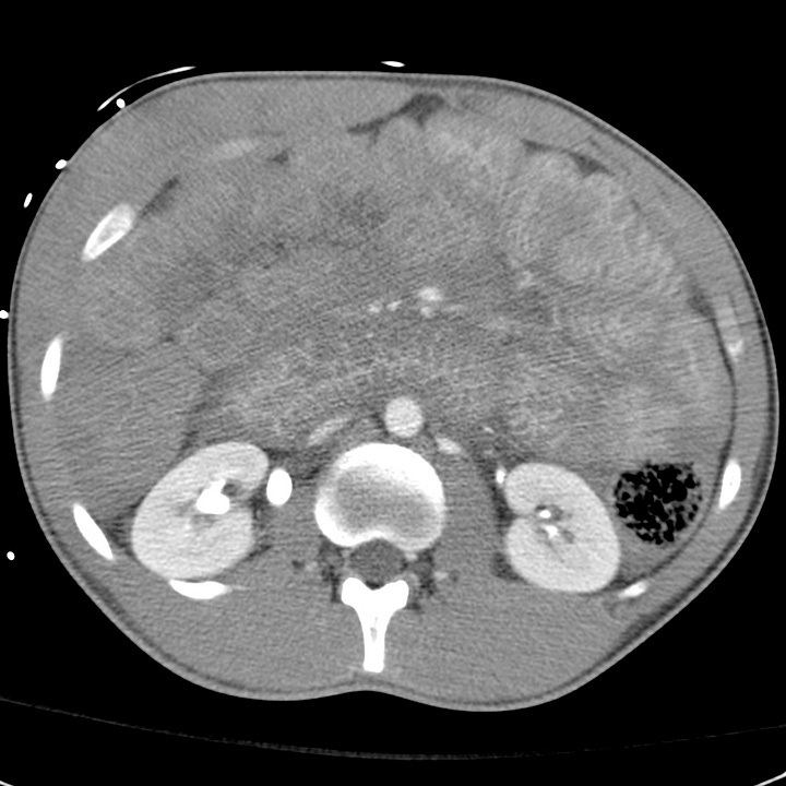
Shock bowel (two patients). The small bowel wall is diffusely edematous with dramatic mucosal enhancement, most pronounced in the proximal small bowel. In both cases, the inferior vena cava is flattened and the kidneys enhance intensely into the excretory phase. Intraperitoneal and intramesenteric fluid are present.
Small-bowel injuries are often clinically subtle. Distracting injuries, intoxication, and depressed consciousness limit the sen- sitivity of physical examination. Small amounts of intraperitoneal fluid may be missed on initial FAST examination, and peritoneal signs may be delayed. Witnessed or suspected abdominal impact and abdominal wall bruising should raise suspicion for bowel contusion or other gastrointestinal injury.
CT has decreased the historic need for exploratory laparotomies after blunt abdominal trauma and should be obtained in any stable patient. Focal small-bowel thickening indicates a probable contusion, and may be associated with discrete intramural or mesenteric hematoma.
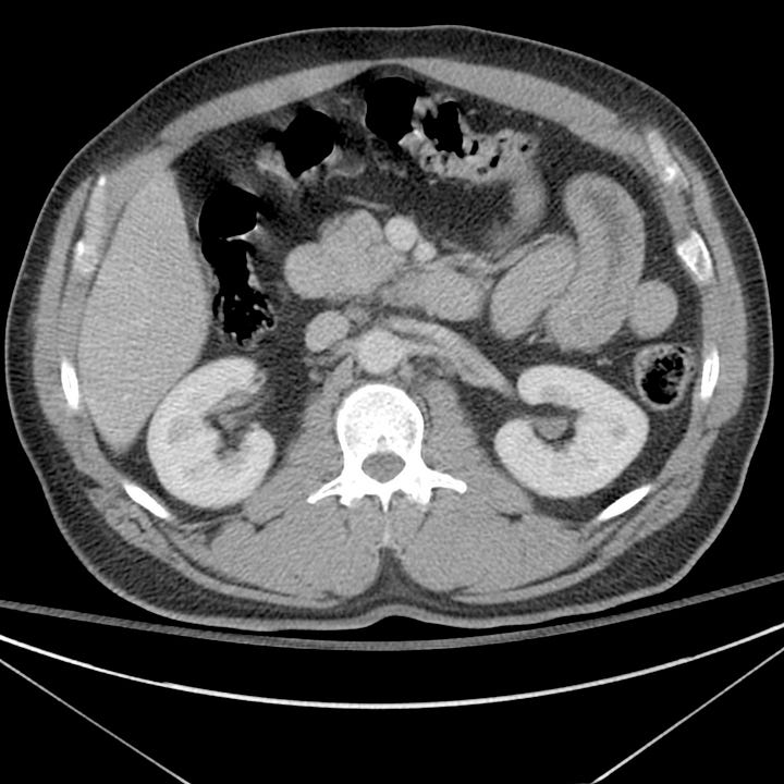
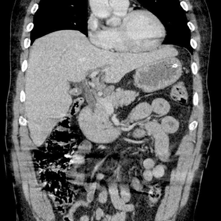
Blunt small-bowel injury. Thickened proximal small-bowel loops with the appearance of “Cheerios” in transverse section.
The consequences of small-bowel laceration are blood loss and peritoneal contamination. Spillage of the intestinal contents into the abdomen ultimately produces a suppurative peritonitis that may take up to 8 hours to manifest clinically, or longer if the patient is unconscious or sedated. Concomitant mesenteric and visceral injuries are often present.
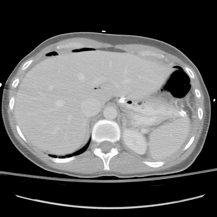
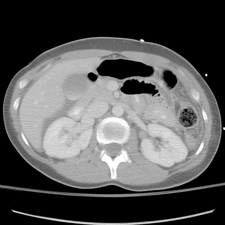
Small-bowel laceration. Free intraperitoneal air and fluid interposed between the duodenum and gallbladder.
Pneumoperitoneum, oral contrast extravasation, bowel wall discontinuity, or mesenteric vascular extravasation are indications for urgent operative exploration. In contrast to most solid organ injuries (e.g., spleen or liver), which can often be safely observed, bowel perforation must be managed surgically.
Small amounts of free fluid, bowel wall thickening, mesenteric stranding, and bowel mucosal enhancement are suggestive findings, and surgery should be considered if two or more of these are present. In penetrating trauma, such as stab wounds, small amounts of extraluminal gas are nonspecific and can be due to air from the wound tract.


