Anterior shoulder dislocation
Anterior shoulder dislocation is the most common type of shoulder dislocation (~ 95%) and occurs with forced arm abduction, external rotation, and extension. The humeral head is displaced anterior, medial, and inferior to its normal location, and its posterolateral surface strikes the anteroinferior surface of the scapular glenoid.
Pain and muscle spasm are the rule, and patients typically hold the affected arm in slight abduction and external rotation.
Anterior shoulder dislocations are well characterized by standard radiographic series consisting of AP views in internal and external rotation, the scapular-y view, and the axillary view. CT or MR may be useful for detection of subtle osteocartilaginous fractures or intra-articular fragments. MR, in particular, is superior for identification of rotator cuff, capsular, and glenoid labral injuries.
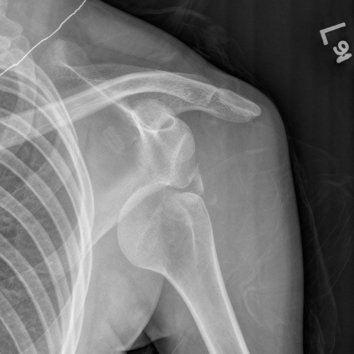
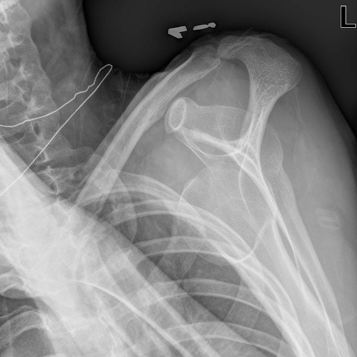
Anterior shoulder dislocation. The humeral head is dislocated inferiorly and anteriorly with respect to the scapular glenoid. On the scapular “Y” view the humeral head is normally centered over the “Y" made up of the junction of the scapular body and spine. In this case it is anteriorly displaced. Several small bony fragments adjacent to the glenoid reflect an associated bony Bankart lesion.
In anterior dislocation, impaction fractures of the posterolateral humeral head (Hill-Sachs lesion) and anteroinferior glenoid labrum avulsion (Bankart lesion) commonly occur. An osseous glenoid rim fracture, when present, is referred to as a bony Bankart lesion.
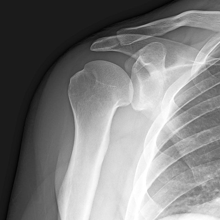
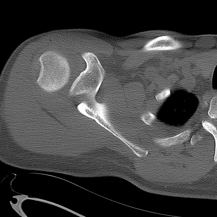
Hill-Sachs lesion. Radiograph and CT after reduction of anterior shoulder dislocation; lateral humeral head impaction fracture.
Luxatio erecta
Luxatio erecta (inferior dislocation) is an uncommon anterior dislocation variant that tends to occur in elderly individuals. It results from forceful hyperabduction with impingement of the humeral neck on the acromion. The humeral head is inferiorly displaced with the humeral shaft directed superolaterally along the glenoid margin. The humeral articular surface is directed inferiorly and is no longer in contact with the inferior glenoid rim.
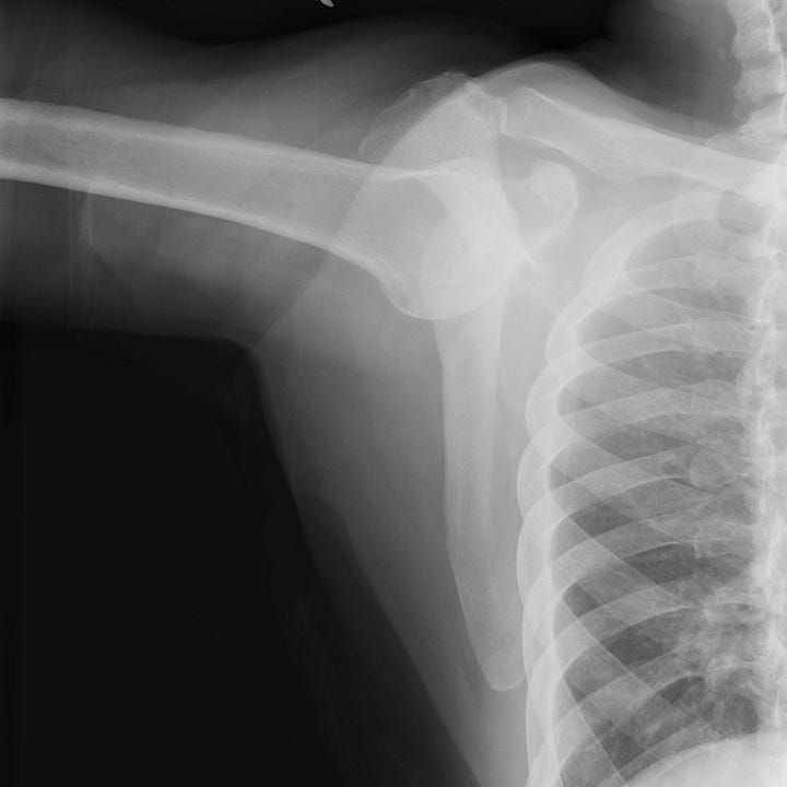
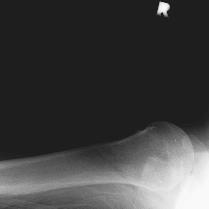
Luxatio erecta. Anterior-inferior right humeral head dislocation with fixed adduction of the humerus.
Luxatio erecta is almost always accompanied by detachment of the rotator cuff and neurovascular compression. Associated fractures are also common. Early reduction should be attempted in an effort to prevent neurologic or vascular damage.
Posterior shoulder dislocation
Posterior shoulder dislocation is much less common than anterior dislocation (~ 5%) and can be a difficult diagnosis both clinically and radiographically. Injury mechanisms include seizures, electrocution, or a blow to the back of the shoulder with the arm internally rotated and abducted. Patients are usually unable to rotate their arm externally, and these injuries can be missed if axillary or scapular “Y” views are not obtained.
On frontal radiographs, the normal slight superposition of the medial humeral head on the glenoid is lost. Because patients cannot externally rotate the affected arm, only internally rotated AP radiographs are possible; these do not show the greater tuberosity in profile and the humeral head looks like a light bulb. When the AP external rotation view appears identical to the internal rotation view, consider posterior dislocation!
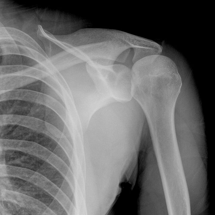
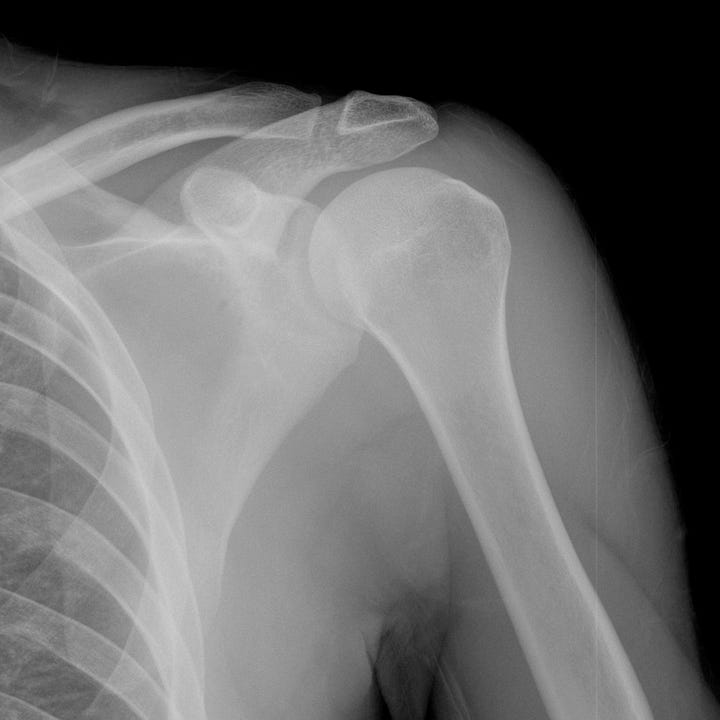
Posterior shoulder dislocation. Frontal radiograph shows lateral displacement of the humeral head with little overlap of the glenoid fossa. The humeral head has a typical “light bulb” appearance with poor visualization of the greater tuberosity. Postreduction radiograph for comparison; the humerus now articulates normally with the glenoid.
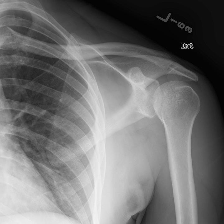
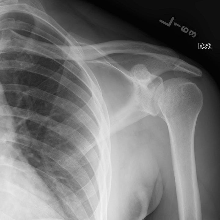
Subtle posterior shoulder dislocation. The orientation of the humeral head is unchanged on internal and external rotation radiographs and demonstrates the “lightbulb” sign. A subtle, vertically oriented crescentic lucency is superimposed on the mid-humeral head (trough sign).
A fracture of the anterior humeral head, known as the trough sign or reverse Hill- Sachs lesion, reflects impaction of the anterior humeral head against the posterior glenoid rim. The corresponding fracture of the posterior glenoid (when present) is called a reverse Bankart lesion.
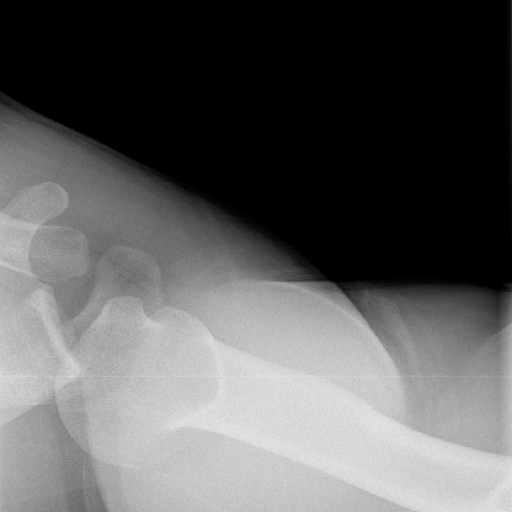
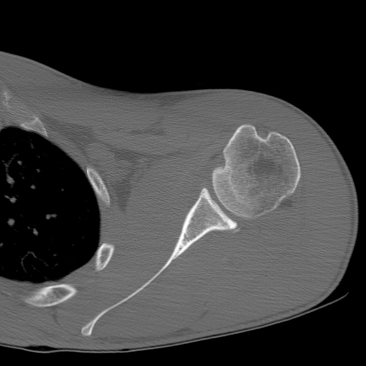
Reverse Hill Sachs and Bankart lesions. Axillary view shows superposition of the humeral head on the posterior glenoid. Postreduction axial CT shows impaction at 10 o’clock, consistent with a reverse Hill-Sachs lesion, and a tiny avulsion of posterior glenoid; a reverse Bankart lesion.


Lex Erecto is my porn name.