Scaphoid fracture is usually due to a fall on an outstretched hand and is the most commonly fractured carpal bone. Patients present with wrist pain and tenderness over the “anatomic snuffbox,” the dorso-radial aspect of the wrist near the base of the thumb. The standard radiographic series (PA, lateral and oblique) may not be diagnostic and a supplemental scaphoid view, a frontal radiograph with the wrist in ulnar deviation, should always be obtained if scaphoid injury is suspected. CT and MRI are highly sensitive and can detect subtle fractures and bone contusions in the patient with negative wrist radiographs. Radiographs obtained 1 to 2 weeks after the injury will also sometimes reveal a previously occult fracture.
Scaphoid fractures can be located at the distal pole (10%), waist (70%), or proximal pole (20%). The primary vascular supply to the scaphoid is via the radial artery, two branches of which supply the distal pole and waist of the scaphoid. With no direct arterial supply, the proximal pole depends on fracture union for revascularization. Delayed diagnosis or nonunited fracture may lead to proximal scaphoid avascular necrosis, which appears as sclerosis, fragmentation, and collapse.
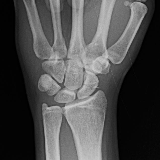
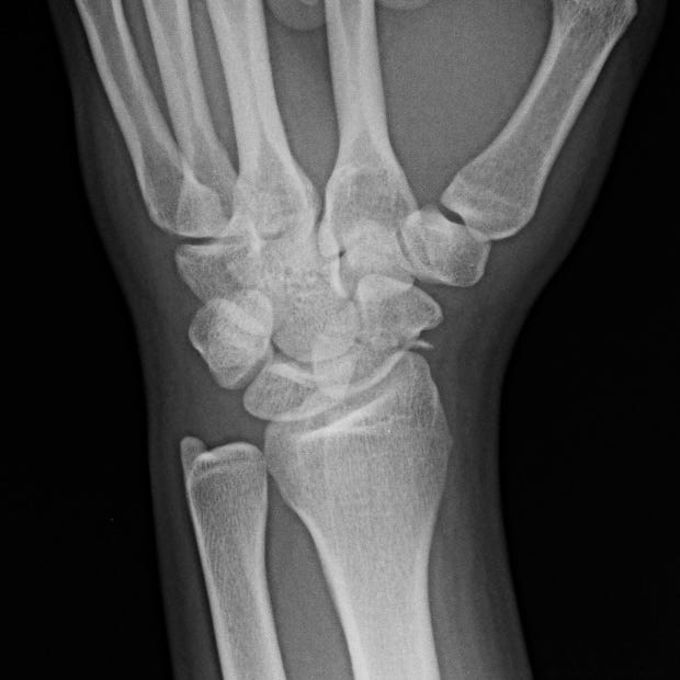
Scaphoid waist fracture. Acute, nondisplaced, minimally comminuted, transverse scaphoid waist fracture.
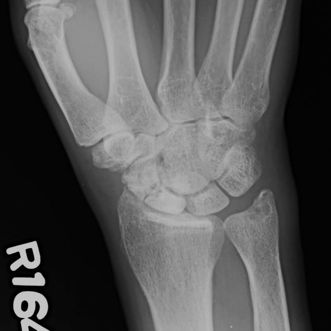
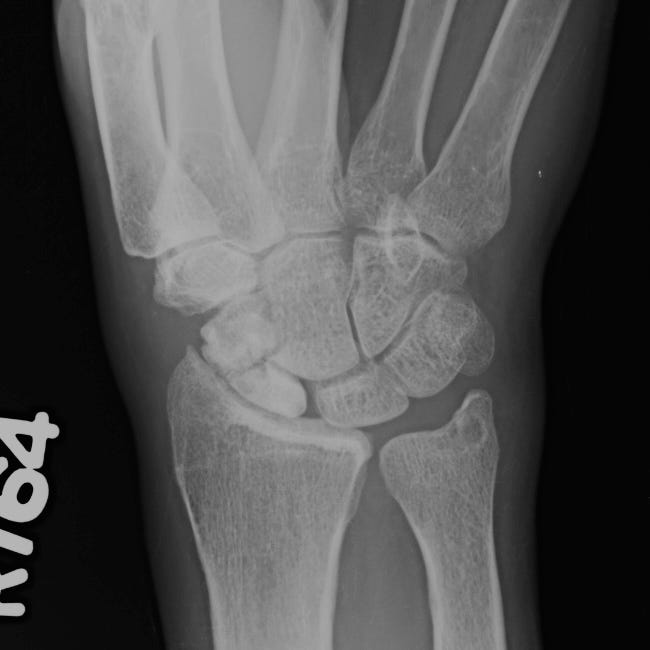
Scaphoid avascular necrosis. Remote, nonunited, scaphoid waist fracture with dense sclerosis and fragmentation of the proximal and distal fragments.
Primary management is nonoperative with wrist immobilization in slight flexion and radial deviation. Time to union can vary based on location of the fracture and may be as long as 24 weeks for proximal scaphoid fractures. Surgical intervention is reserved for nonunited, displaced, or unstable fractures.
Scapholunate dissociation is the most common ligamentous disruption of the carpus and is often associated with scaphoid fracture. In trauma, the scapholunate ligament fails under high-energy loading of the extended, ulnar-deviated wrist. Scapholunate dissociation can also be seen in the setting of chronic arthritis. On PA radiographs, the normal scapholunate interval is less than 2 mm. In scapholunate dissociation it is greater than 3 mm. Pain is localized over the dorsal scapholunate region and exacerbated by dorsiflexion. Scapholunate ligament reconstruction may be required to prevent persistent instability.
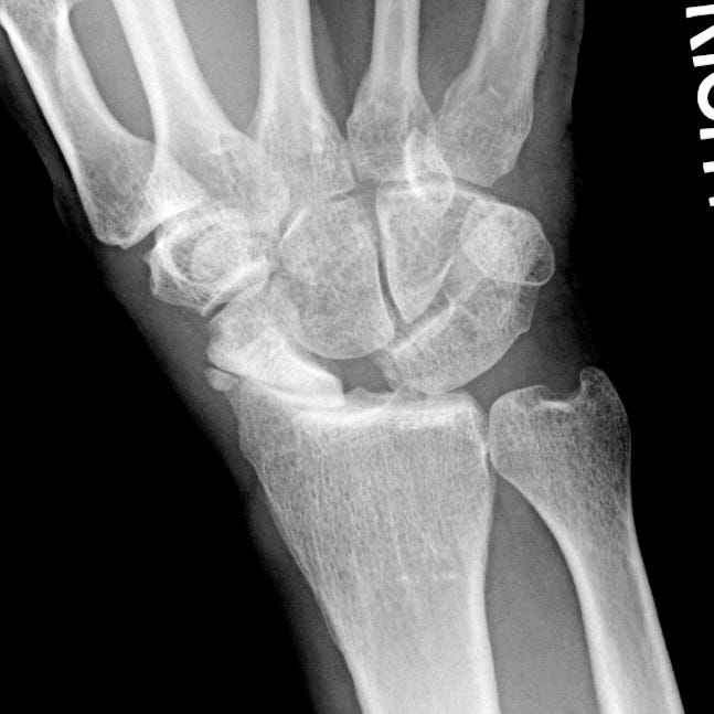
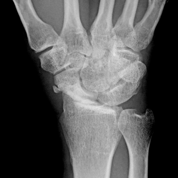
Scapholunate dissociation with remote scaphoid fracture and avascular necrosis. The scapholunate interval is 6 mm. The proximal scaphoid pole is sclerotic with slight fragmentation at the scaphoid waist. Marked radiocarpal narrowing and subchondral sclerosis.

