Most renal injuries may be due to blunt trauma, often with other associated visceral or spinal injuries, or they may result from penetrating trauma such as a stab wound to the flank. CT identifies subcapsular and parenchymal hematomas (contusion), renal lacerations, arterial extravasation, pseudoaneurysms, and collecting system injury. If a renal abnormality is found on initial workstation review, delayed images through the kidneys and collecting system should be obtained to detect arterial extravasation, pseudoaneurysm, or urine extravasation.
The primary aim in evaluating acute renal trauma is prevention of life-threatening hemorrhage by promptly identifying arterial bleeding and/or pseudoaneurysm, which can be managed by embolization, or in severe cases, by surgery.
Renal contusions are poorly defined, round, or ovoid low-attenuation areas and should be distinguished from renal infarcts, which are sharply delineated, usually peripheral, and wedge-shaped. Subcapsular hematomas are elliptical hemorrhages interposed between the renal capsule and the kidney parenchyma
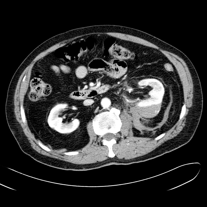
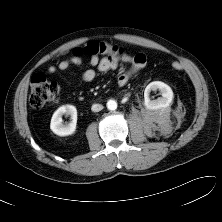
Subcapsular hematoma (grade II). Left subcapsular and inferior posterior perirenal hematoma confined to the retroperitoneum. No parenchymal laceration.
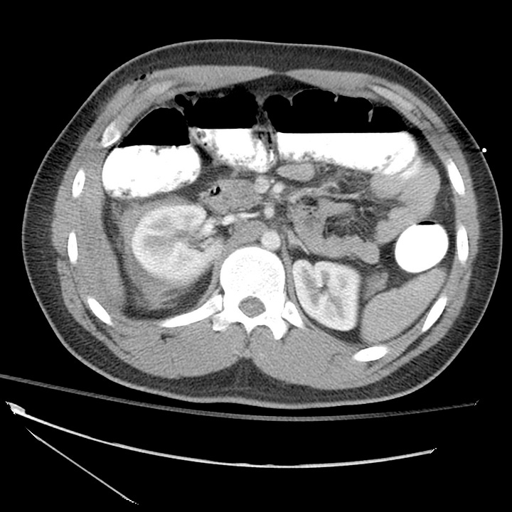
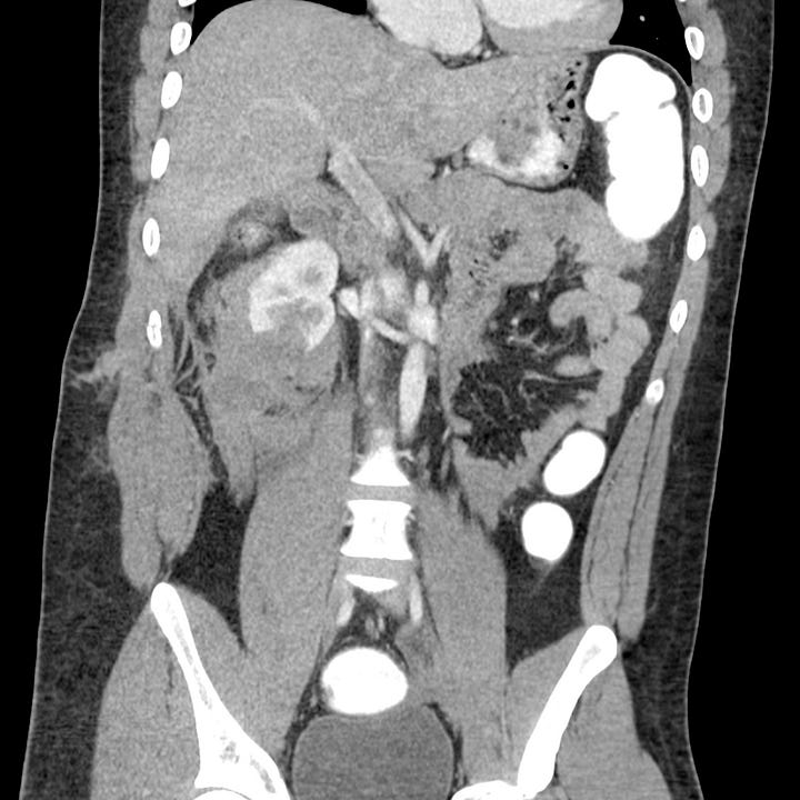
Renal laceration (grade III). Stab wound to right flank with 2-cm inferior renal laceration and retroperitoneal hematoma. No active arterial extravasation or pseudoaneurysm.
Major injuries comprise approximately 10% of renal injuries and include lacerations that extend from the cortex to the collecting system, multiple renal lacerations (known as shattered kidney), segmental infarcts, and vascular injury to the renal pedicle (often with active hemorrhage).
Sudden deceleration can disrupt the ureteropelvic or ureterovesical junctions. Surgical mishap, ureteroscopy, gunshot, and penetrating trauma are other causes. Collecting system disruption is confirmed on delayed images by identifying contrast medial to the kidney or along the course of the ureter. Collecting system injuries are classified as complete (avulsion) or incomplete (laceration). Consequences of delayed diagnosis include urinoma, abscess, and stricture.
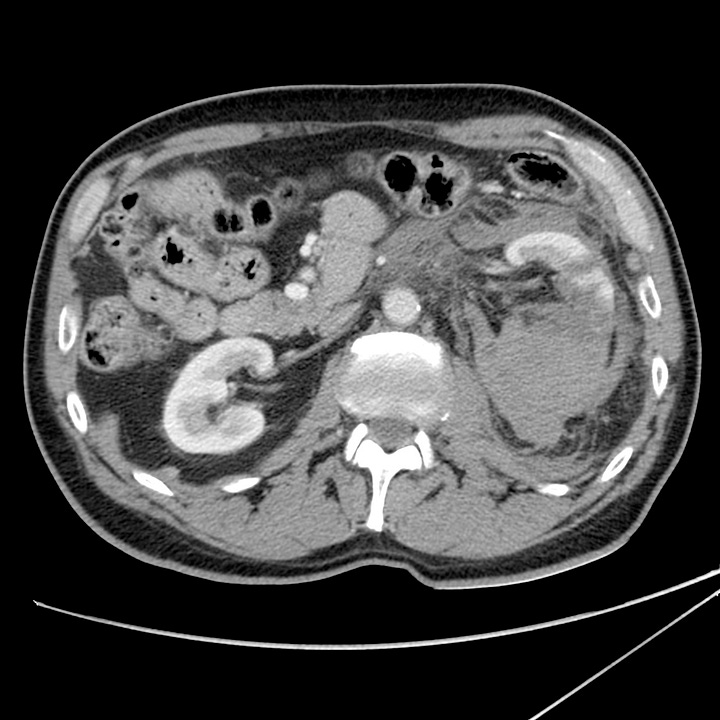
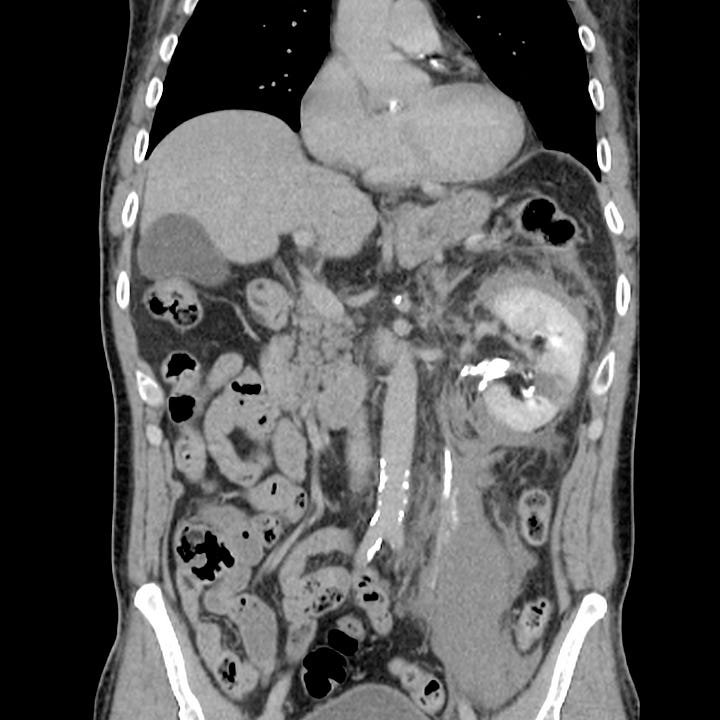
Collecting system disruption (grade IV–V). Initial axial image shows a large parenchymal hematoma. Delayed coronal image shows contrast and urine extravasation inferomedial to the kidney.
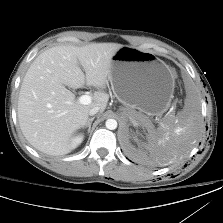
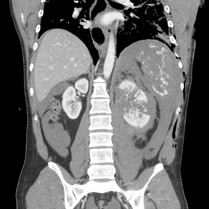
Shattered kidney and spleen (grade V). Blunt trauma. Shattered kidney and spleen with extensive hemoperitoneum and active arterial contrast extravasation.
Renal Injury Grading
Grade I
Hematuria with normal kidney, renal contusion or nonexpanding subcapsular hematoma.
Grade II
Nonexpanding perirenal hematoma, confined to the retroperitoneum. Laceration < 1 cm in depth, no urinary extravasation.
Grade III
Laceration > 1 cm parenchymal depth, without collecting system rupture or urinary extravasation.
Grade IV
Laceration through renal cortex, medulla, and collecting system; renal artery or vein injury with contained hemorrhage.
Grade V
Completely shattered kidney; renal hilum avulsion with devascularized kidney
All grade I and II injuries and 90% of grade III and IV injuries are managed con- servatively. Grade V injuries usually require nephrectomy.


