Intracranial air is always seen after craniotomy and also occurs in a small percentage of head trauma patients, usually when a skull base or calvarial fracture traverses one of the facial sinuses or the mastoid air cells. In this case air may be seen in the epidural, subdural, or subarachnoid space. Air within venous structures is usually due to air introduced through an intravenous catheter.
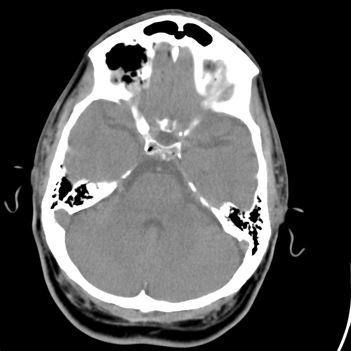
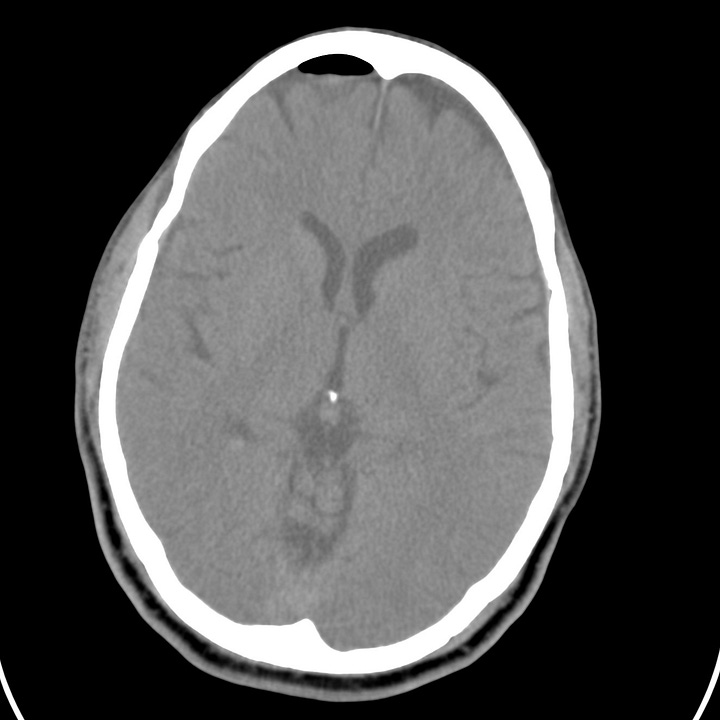
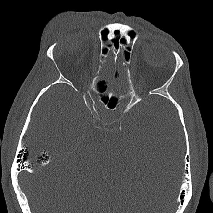
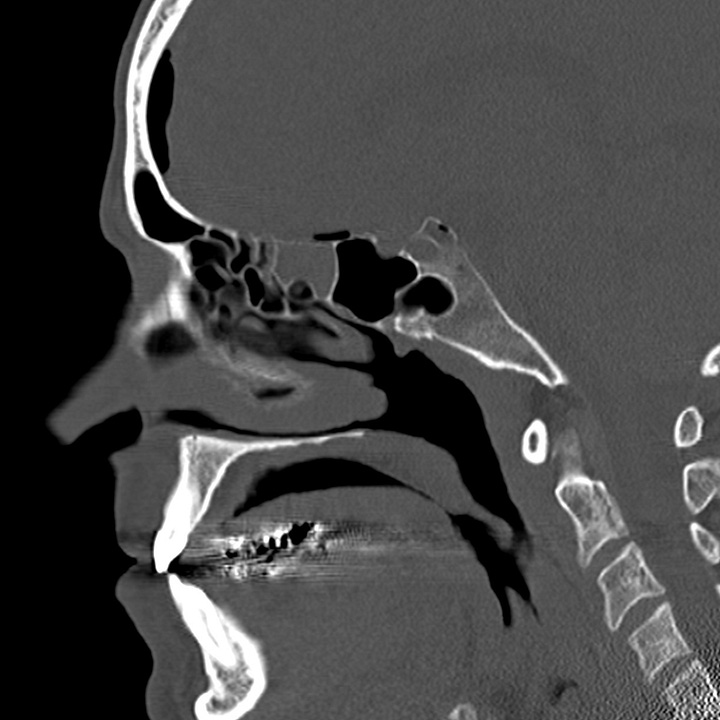
Pneumocephalus following facial trauma with orbital roof and ethmoid fractures. Small amount of air anterior to the right frontal lobe, between the ethmoid bone and the olfactory cortex, and within the right superior orbit. This air is not under tension, but it indicates that the skull base periosteum has been breached and that the subdural space communicates with one of the facial sinuses (in this case the ethmoid air cells). The patient is therefore at increased risk of CSF leak or meningitis.
Rarely, a dural laceration can act like a one-way check valve, permitting inflow of air, but preventing its egress. By this mechanism, intracranial air pressure can exceed tissue pressure and compress the brain. In the supine patient, this is most evident in the anterior cranial fossa and results in a peaked appearance of the frontal lobes (Mount Fuji sign).
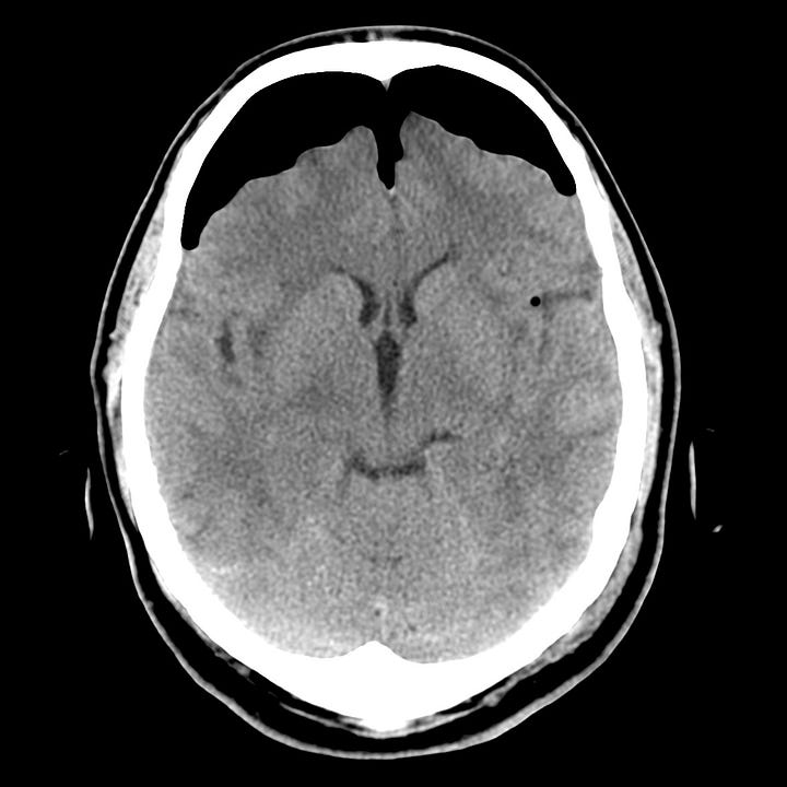
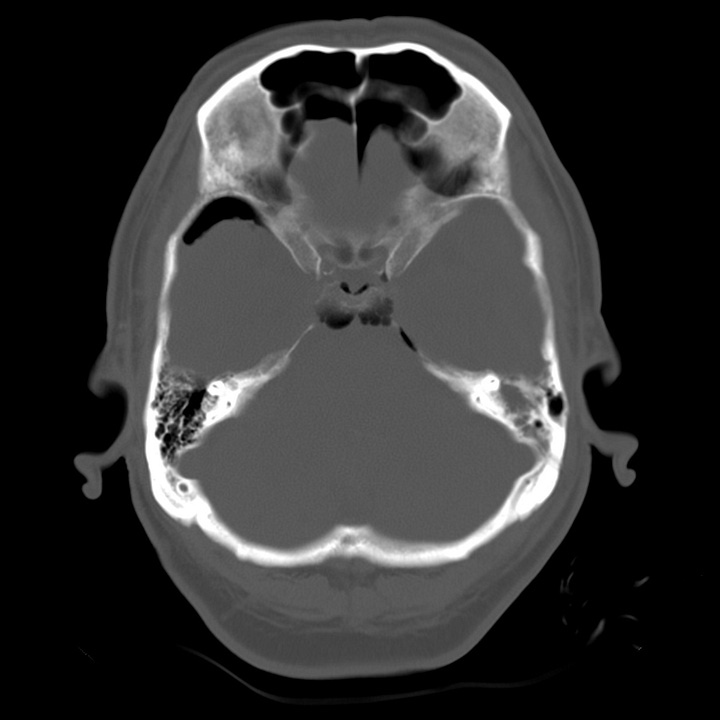
Tension pneumocephalus due to temporal bone fracture. Bifrontal subdural air collections with mild compression of the frontal parenchyma. Opacified left mastoid air cells are consistent with an acute tem- poral bone fracture. Air in the cisterns and left sylvian fissure indicate arachnoid injury.
Most cases resolve spontaneously and can be observed. Antimicrobial therapy has been employed to prevent meningitis when a disrupted sinus or mastoid air cell communicates with the intracranial cavity, but prophylaxis is controversial and is usually only indicated in the presence of CSF leak. If associated with neurologic deterioration, tension pneumocephalus can be treated with burr hole decompression.

