Orbital wall fractures usually result from an abrupt increase in intraorbital pressure, usually when a fist or ball strikes the eye. The thin medial or inferior orbital walls break with variable degrees of orbital fat herniation, extra-ocular muscle entrapment, orbital emphysema, and intrasinus hemorrhage. These are often referred to as orbital blowout fractures.
Clinical findings in orbital floor fractures include restricted upward and lateral gaze, subcutaneous emphysema, and diminished sensation in the distribution of the infraorbital nerve (V2). Enophthalmos is usually not immediately evident, but it can be seen in unrepaired fractures after initial swelling resolves. Rarely symptomatic bradycardia can result from stretching of the infraorbital nerve (oculocardiac reflex).
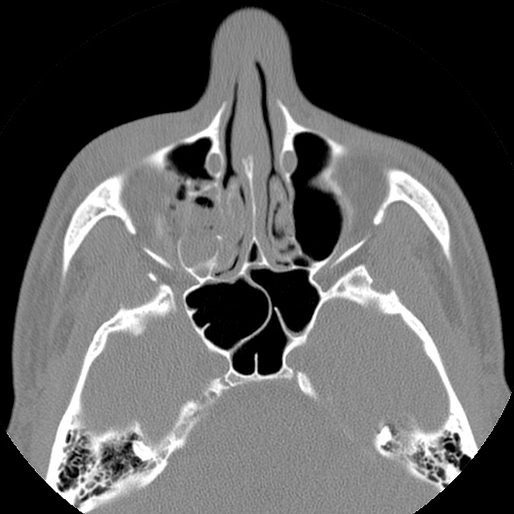
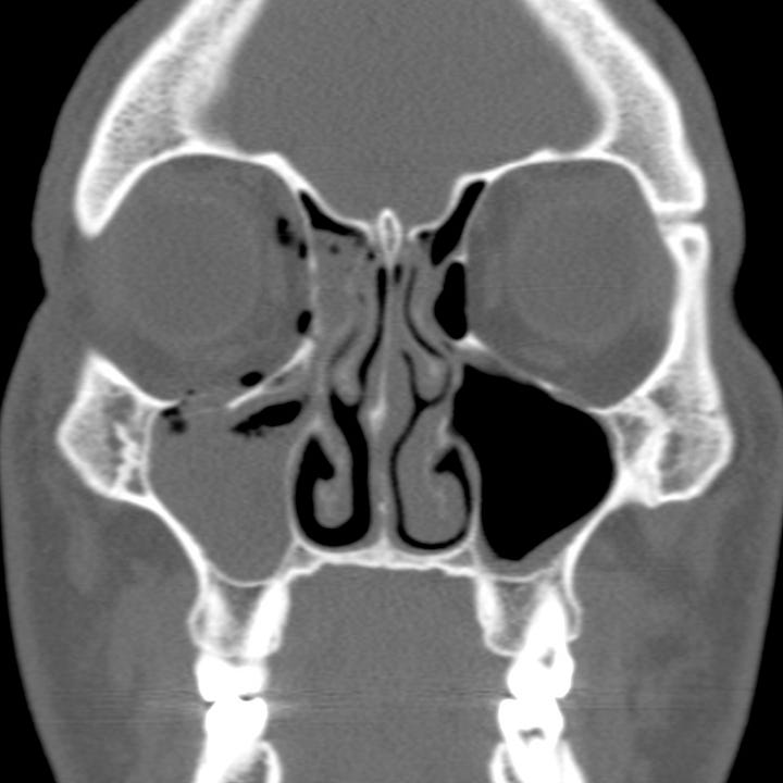
Acute orbital floor fracture. Right orbital floor fracture with depression of the lateral orbital floor. The infraorbital canal (V2 branch) is intact. Intraorbital air and intrasinus hemorrhage.sts and
While medial orbital wall (lamina papyracea) fractures often occur in conjunction with floor fractures, isolated medial wall fractures are uncommon. Most are small, of little consequence, and discovered on CT obtained for other indications long after an injury. When symptomatic, medial wall fractures are associated with orbital emphysema and potential medial rectus muscle entrapment, which can lead to diplopia on lateral gaze.
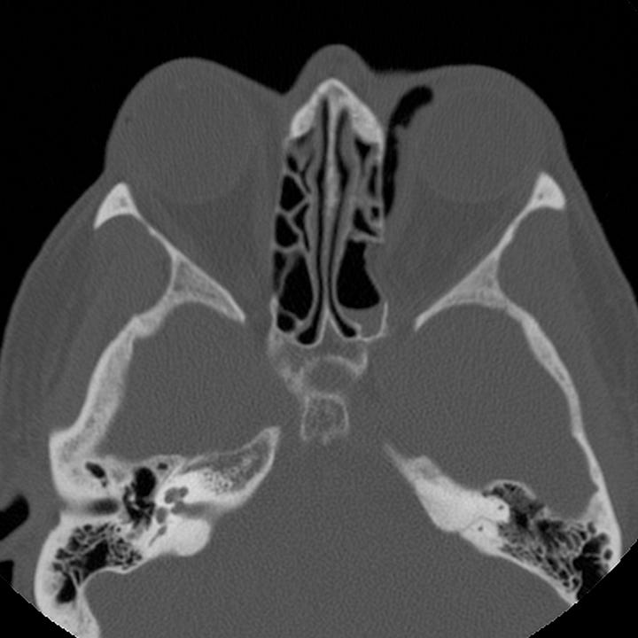
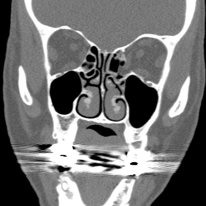
Acute medial orbital wall fracture. Left posterior medial orbital wall fracture with intraorbital emphysema and fat and medial rectus herniation into the posterior ethmoid air cells. The medial rectus is tethered at the anterior margin of the fracture.
Orbital roof fractures are also uncommon, and may result from direct orbital impact, or more commonly, extension from frontal and calvarial fractures in major head injury.
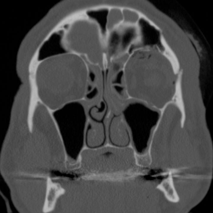
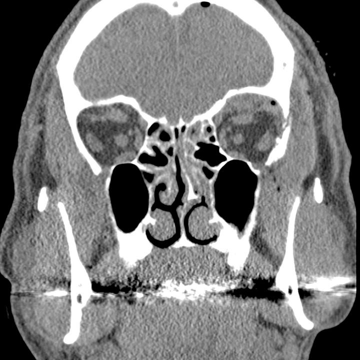
Orbital roof fracture. Left orbital roof “blow up” fracture with extension into aerated frontal sinus. Superior orbital extraconal hematoma and orbital emphysema.
CT defines the area and location of the fracture as well as any fragment displacement. Orbital fat herniation, extra-ocular muscle entrapment, and infraorbital canal involvement are easily identified. Muscle entrapment is evident clinically by diplopia on horizontal or vertical gaze, and corresponding CT findings include an acute change in the angle of the muscle as it passes through the orbit or impalement of one of the muscles on a bone spicule. Intraorbital emphysema is associated with risk of infection and subperiosteal or intraorbital hematoma can result in elevated intra-orbital pressure and secondary globe ischemia or optic nerve damage.
Surgical intervention is usually indicated for severe fractures to prevent late enophthalmos and diplopia. Emergent surgical indications include symptomatic bradycardia and large orbital hematoma.


