Olecranon fractures are common, usually intra-articular, and typically seen in older adults who sustain either direct impact to the elbow or forced hyperextention from standing height falls. Associated distal humeral, radial head and coronoid process fractures, if present, may result in an unstable “floating” elbow. Because the triceps inserts on the olecranon, its unopposed action often results in wide separation of the fragments.
Clinical features include pain, swelling and inability to extend the forearm against gravity. Ulnar nerve injury is possible
Best seen on the lateral elbow radiograph, olecranon fractures appear as a defect extending from the dorsal ulnar cortex to the articular surface of the trochlea. Type A fractures ( AO classification) are extra-articular, Type B fractures are intra-articular, and Type C fractures are associated with radial head fractures.
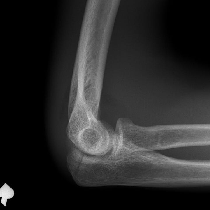
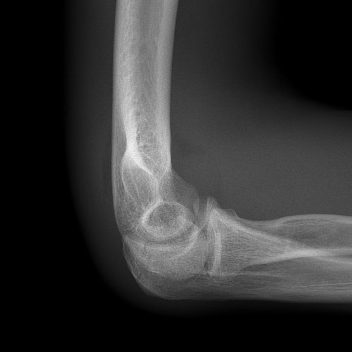
Nondisplaced olecranon fracture (Type A). Subtle, nondisplaced intra-articular fracture.
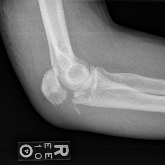
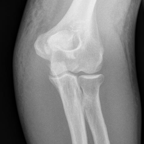
Displaced intra-articular olecranon fracture (Type B). Minimally comminuted intra-articular olecranon fracture with extension to the trochlear joint surface. Approximately 8 mm fragment displacement; marked soft tissue swelling.
In most cases, olecranon fractures require open reduction and internal fixa- tion to restore the articular surface and preserve the elbow extensor mechanism. Nondisplaced fractures can be managed conservatively. Post-traumatic arthritis occurs in ~ 20% of patients, particularly if reduction is suboptimal.
Triceps avulsion injuries are unusual and are characterized on radiographs by a small, proximally displaced osseous flake or enthesophyte fragment. Usually the result of a fall on an outstretched hand with the elbow in flexion, triceps avulsion injuries may be seen in weightlifters, patients with renal disease, and patients who receive systemic steroid therapy or local steroid injection.
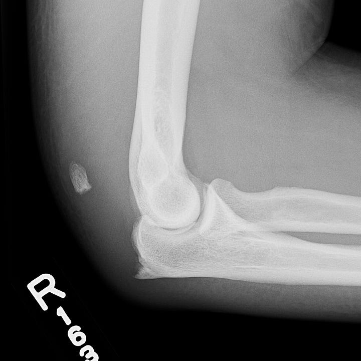

Triceps avulsion. A fractured enthesophyte at the triceps insertion is proximally displaced with respect to the olecranon.


