Odontoid (dens) fracture
Spine
C2 odontoid fractures are usually low energy injuries seen in elderly patients who fall from standing or sitting. In younger patients, odontoid fractures are typically associated with higher energy mechanism head injuries. Hyperflexion, hyperextension, and lateral flexion can all result in fractures that separate the odontoid from the C2 vertebral body. CT is the standard imaging modality for acute cervical spine trauma in adults, although radiographs are usually appropriate in young children and may be obtained when CT is not available. Radiographs, if obtained, should include both lateral and AP odontoid views.
Type I fractures are avulsions of the odontoid tip at the attachment of the alar liga- ments. These are rare but sometimes seen in patients with atlantooccipital dislocation. Type II fractures traverse the odontoid base. Type III fractures separate a fragment that includes both the dens and a portion of the C2 body.
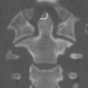
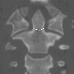
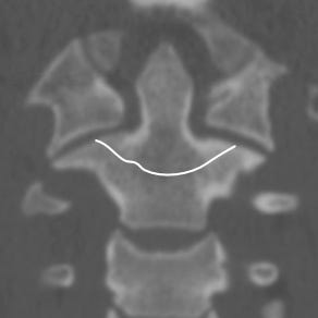
Odontoid fracture classification. Type I fractures are odontoid tip avulsions at the attachment of the alar ligament. Type II fractures traverse the base of the dens. Type III fractures involve only the C2 body.
Because the dens is mainly composed of dense cortical bone, type I and II fractures are prone to non-union, and patients often require C1-C2 fusion or anterior odontoid screw fixation. Thanks to the larger area of cancellous bone-to-bone contact, type III fractures tend to heal with immobilization and do not usually require surgical fixation. Type II and III fractures are also referred to as “high” and “low” dens fractures, respectively.
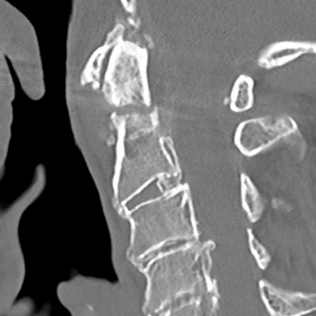

Type II odontoid fracture. Transverse fracture through the base of the odontoid with adjacent epidural and prevertebral hematoma.
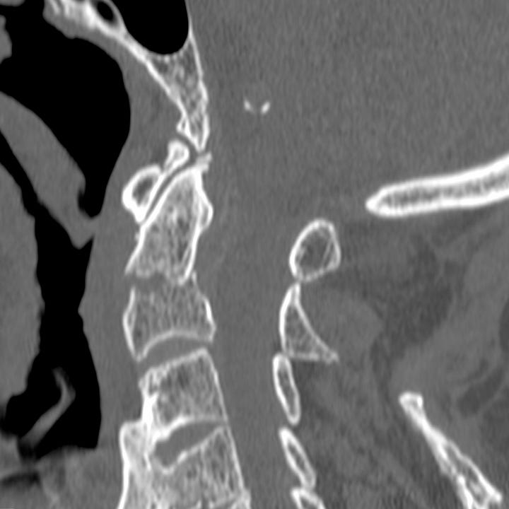
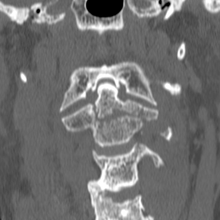
Type III odontoid fracture. Transverse fracture through the upper body of C2 with minimal dorsal angulation of the odontoid fragment.

