On CT, cerebral edema is lower in density than surrounding normal parenchyma. On MRI, signal intensity of edema is lower than that of brain parenchyma on T1-weighted images and higher on T2-weighted images. Diffusion-weighted images (DWI) can distinguish between cytotoxic edema (restricted diffusion) and vasogenic or interstitial edema (normal or increased diffusion). Fluid attenuation inversion recovery (FLAIR) sequences suppress intraventricular CSF signal and are sensitive for detecting adjacent areas of interstitial edema (Fig. 2.3).
Localized cerebral edema may be classified as cytotoxic, vasogenic, or interstitial, although overlap is often present. Cytotoxic edema refers to intracellular swelling caused by depletion of adenosine triphosphate (ATP) and consequent sodium/potassium membrane pump failure. Most commonly seen in cerebral infarct and anoxic injury, cytotoxic edema involves both white and gray matter similarly on most imaging studies.
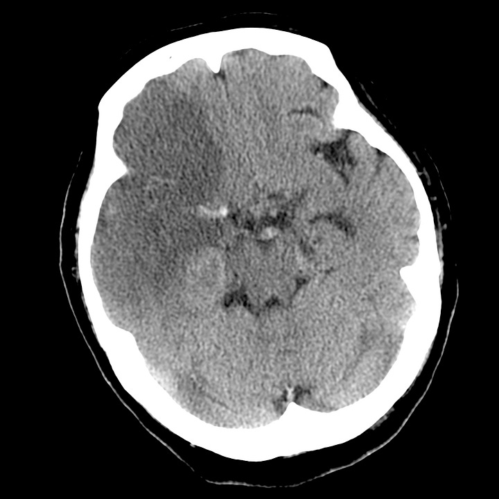

Cytotoxic edema in subacute cerebral infarction. Extensive right frontal and temporal low-density change with absent differentiation between gray and white matter, sulcal effacement, and ipsilateral ventricular compression. The middle cerebral artery is dense, indicating thrombosis.
Vasogenic edema, in contrast, preferentially involves white matter and results from conditions that increase capillary permeability. Vasogenic edema is often due to brain contusion, severe hypertensive disease, primary and metastatic neoplasms,
intracranial abscess or other infectious/inflammatory processes.
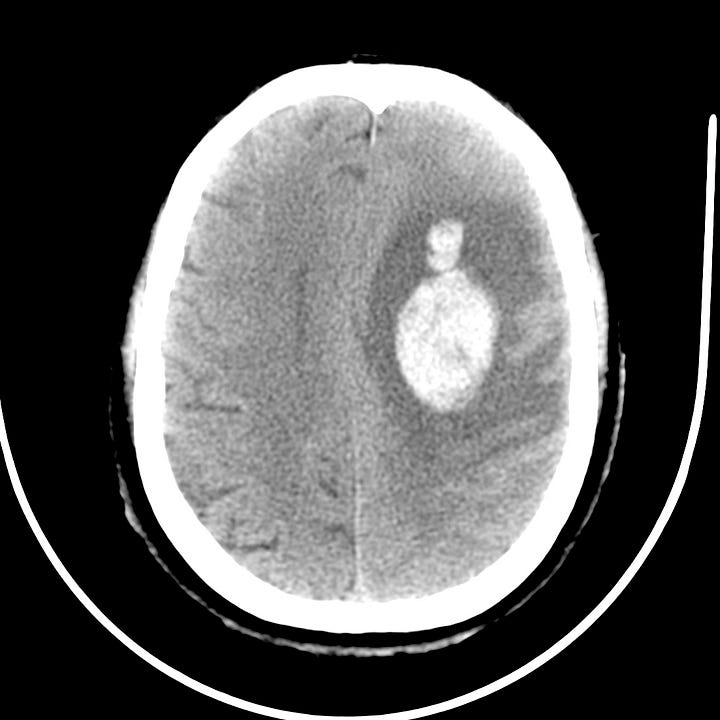
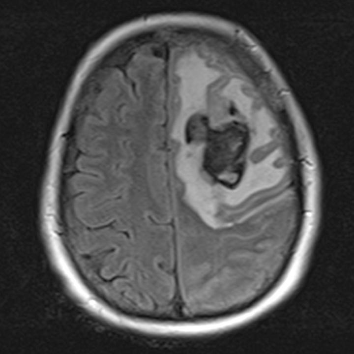
Vasogenic edema due to hemorrhagic brain metastasis from lung carcinoma. Left frontal white matter hemorrhage with surrounding low attenuation edema that spares the cortical gray matter. FLAIR MRI shows a hemorrhagic nodule with surrounding high-signal edema that is limited to the white matter.
Acute hydrocephalus can cause interstitial edema, as elevated intraventricular pressure leads to trans-ependymal migration of CSF into the periventricular white matter, where it is resorbed via parenchymal capillaries.
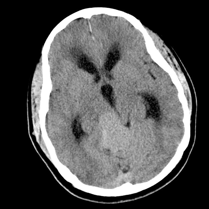
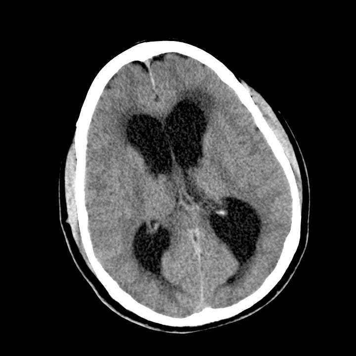
Interstitial edema due to fourth-ventricle obstruction by meningioma. Acute hydrocephalus with low-density trans-ependymal CSF resorption, most conspicuous at the frontal, occipital, and temporal horns of the lateral ventricle.
