Like flexion injuries, cervical spine extension injuries can range from simple sprains to fracture-dislocations with significant neurologic deficit. The injury mechanism is sudden, severe hyperextension, usually due to impact to the head or lower face. In the less severe hyperextension sprain, prevertebral soft tissues are often swollen due to anterior longitudinal spinal ligament injury, but the middle and posterior vertebral columns remain intact and aligned.
Hyperextension with disruption of the anterior, middle, and posterior ligaments can result in severe spinal cord injury from transient dislocation at the time of impact. Prevertebral soft tissue swelling and subtle anterior disk space widening may be the only imaging evidence of this injury.
In hyperextension teardrop fractures, the anterior longitudinal ligament is avulsed and produces a teardrop fracture of the anteroinferior vertebral endplate. The fragment is characteristically (but not always) larger in the vertical than the horizontal dimension. In elderly patients this fracture usually occurs at C2 and is relatively stable. However, in younger patients, hyperextension injuries often involve the lower cervical spine and can be associated with acute central cervical cord syndrome: motor weakness below the level of the lesion that affects the upper extremities more severely than the lower extremities.
A triangular avulsed fragment is generally visible on lateral radiograph or sagittal CT reformation, with associated anterior disk space widening and prevertebral soft tis- sue swelling. Concomitant hyperextension injuries at other levels are not uncommon. In dislocation, MRI optimally evaluates ligamentous injury, intervertebral disk disrup- tion, epidural hematoma, and spinal cord injury. OnT2-weighted images, acute spinal ligament injury is seen as an interruption in the otherwise low signal intensity ligament surrounded by high-signal edema.
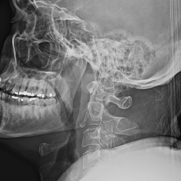
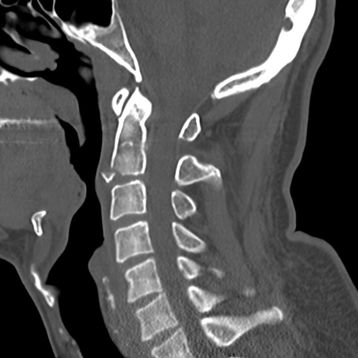
Hyperextension teardrop fracture. C2 anterior inferior avulsed fragment with only minimal associated soft tissue swelling.

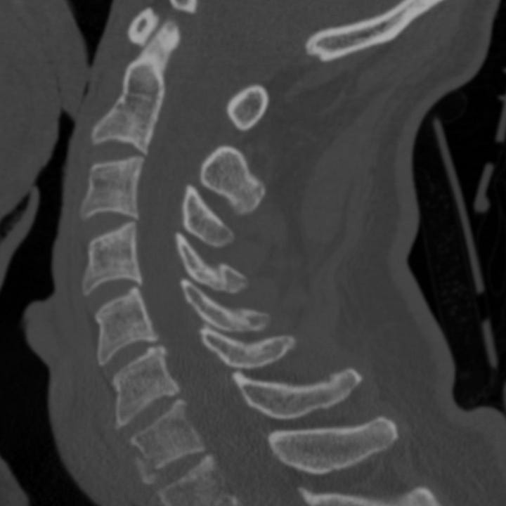
Hyperextension teardrop fracture. C3 avulsion in a patient with a partially fused cervical spine. Moderate prevertebral soft tissue swelling. C5 avulsion fracture in which the small triangular fragment is taller than it is wide (typical).
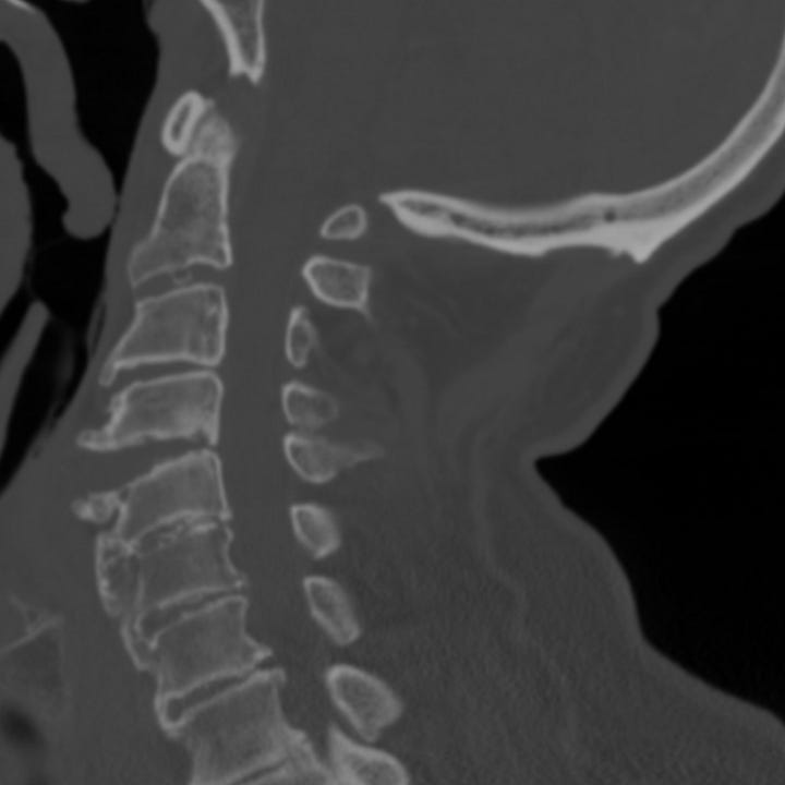
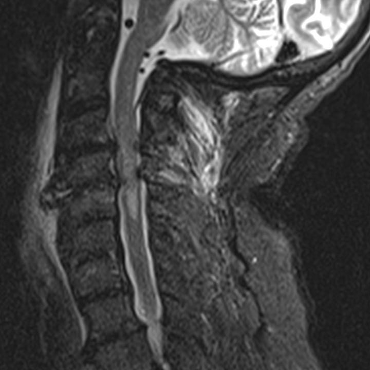
Hyperextension injury without fracture. Isolated widening of anterior C4–C5 disk space. T2-weighted sagittal MRI shows extensive prevertebral soft tissue edema, dorsal nuchal ligament edema, canal stenosis from C3–C4 to C5–C6, and central cord high signal indicating acute spinal injury.


