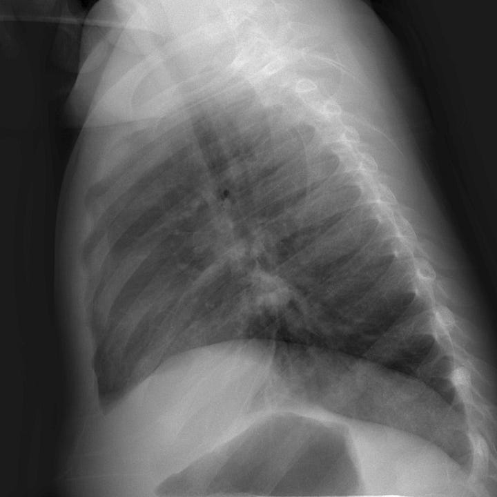
Bronchiolitis is usually due to respiratory syncytial virus (RSV), but rhinovirus, influenza, parainfluenza, and adenovirus are other causes. It is the most common lower respiratory tract infection among children less than 2 years of age, and typically occurs in the late fall to early spring. Affected children have symptoms of cough, and rhinitis. Tachypnea, chest wall retractions, wheezing, and crackles on ascultation may also be present.


Bronchiolitis. Hyperinflation with mildly indistinct central vessels. No dense focal opacity.
Younger infants, ex-premature infants, and infants with congenital heart and lung disease are at higher risk and may present with nonspecific signs of low-grade fever and decreased oral intake.
While most children are managed without imaging. Radiographs are sometimes obtained in the emergency department to exclude pneumonia. When present, imaging findings may overlap with those of acute pneumonia and include peribronchial thickening, hyperinflation, atelectasis, and consolidation.
Treatment consists of supportive care with supplemental oxygen and antipyretics as needed. Antibiotics are not routinely indicated.
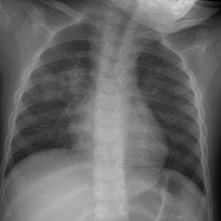
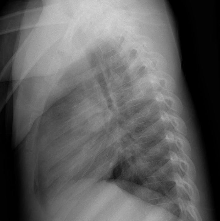
Acute pneumonia in children is usually caused by Streptococcus pneumoniae and appears on plain radiographs as a dense peripheral opacity, sometimes spherical and mass-like (“round” pneumonia). Any focal airspace opacity can be due to pneumonia and should be correlated with supporting clinical features: fever > 102°F, toxic appearance, and leukocytosis.


Right upper lobe pneumonia. Diffuse perihilar opacity visible on both PA and lateral radiographs.
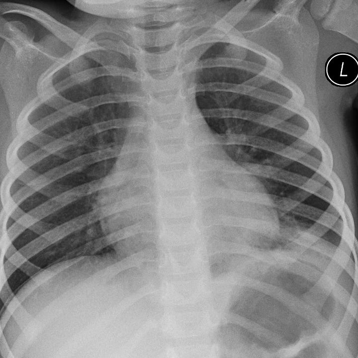
In children, basilar pneumonias can mimic acute abdominal disease such as obstruction or appendicitis. Aspiration pneumonia can be seen in children with neurological conditions that reduce their ability to protect the airway. When aspiration occurs in the supine position, the upper lobes are often involved.


Left lower lobe pneumonia. Dense, mass-like, consolidation with air bronchograms that abuts the diaphragm and is much more conspicuous on the lateral radiograph. Pediatric pneumonia in this location could present clinically as acute abdominal pain.


