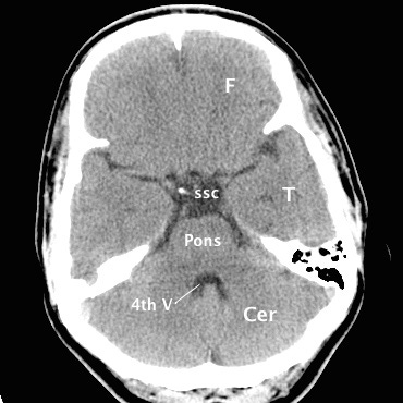
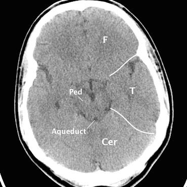

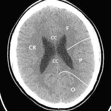
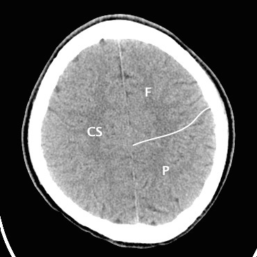
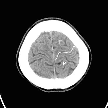
CT Landmarks
F: Frontal lobe P: Parietal lobe T: Temporal lobe O: Occipital lobe Cer: Cerebellum CS: Centrum semiovale CR: Corona radiata CC: Corpus callosum SSC: Suprasellar cistern Th: Thalamus Pu: Putamen C: Caudate head Op: Frontal operculum Ped: Cerebral peduncle
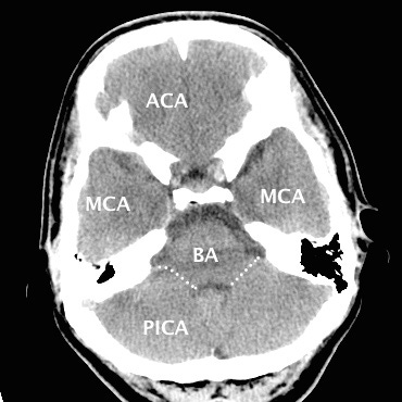
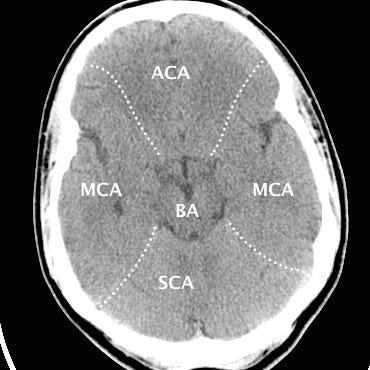
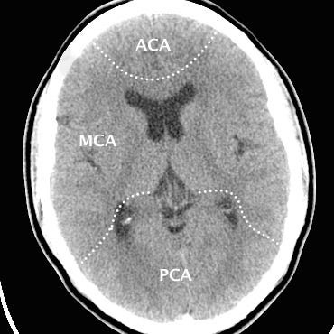

Cerebral vascular territories
ACA: Anterior cerebral artery MCA: Middle cerebral artery PCA: Posterior cerebral artery BA: Basilar artery PICA: Posterior inferior cerebellar artery SCA: Superior cerebellar artery
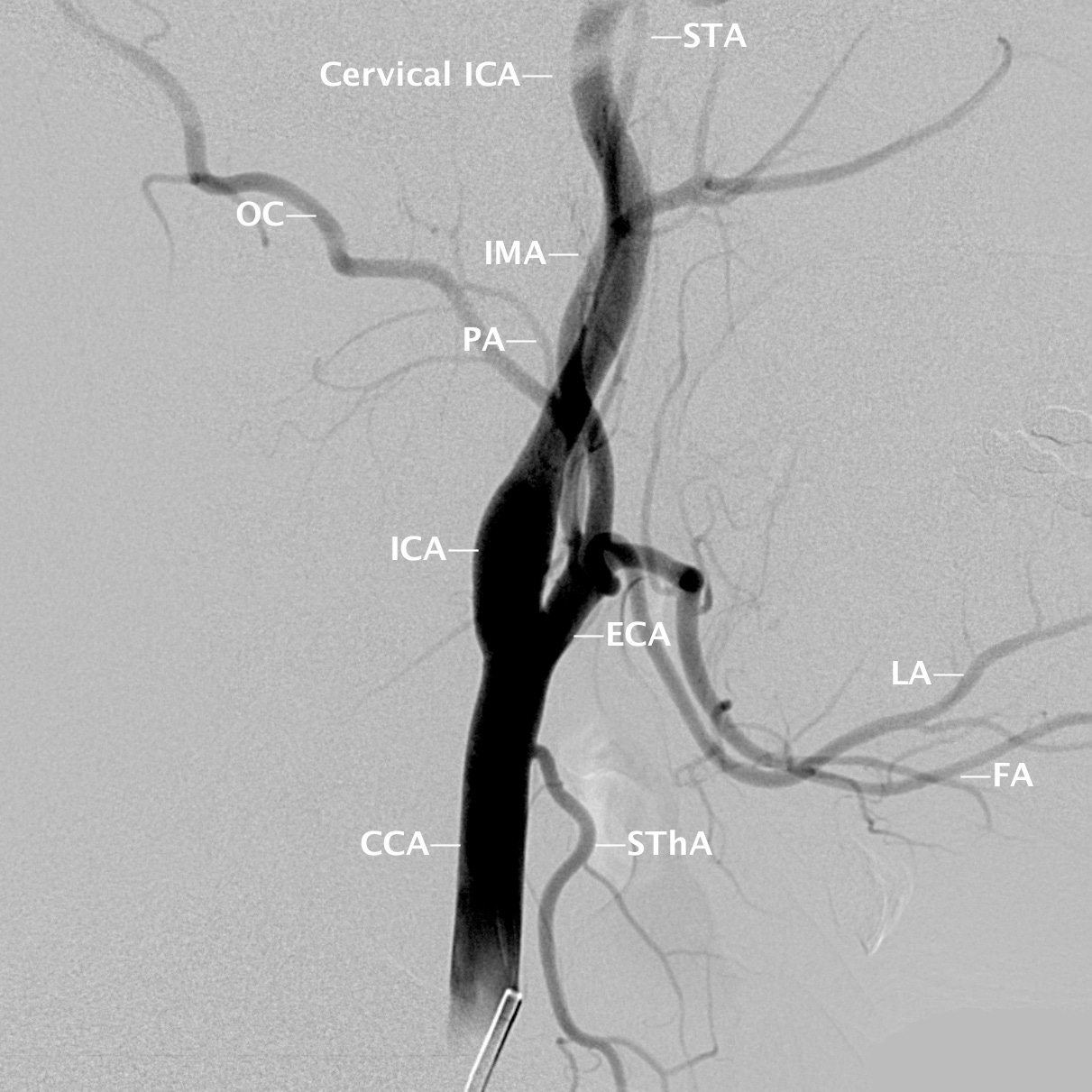
Extracranial carotid anatomy
CCA: Common carotid artery ICA: Internal carotid artery ECA: External carotid artery SThA: Superior thyroidal artery LA: Lingual artery FA: Facial artery PA: Posterior auricular artery OC: Occipital artery IMA: Internal maxillary artery STA: Superficial temporal artery.
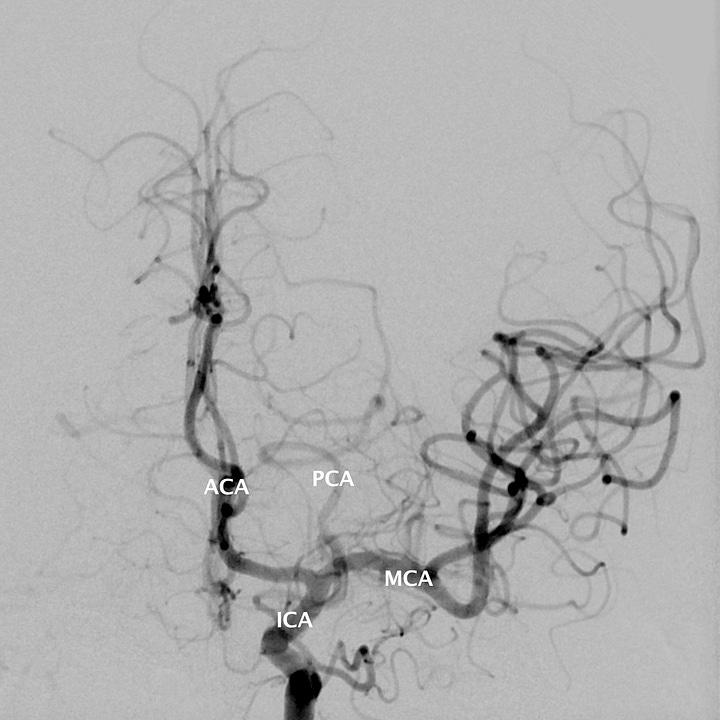

Intracranial carotid anatomy
ICA: Internal carotid artery ACA: Anterior cerebral artery PCA: Posterior cerebral artery MCA: Middle cerebral artery Ach: Anterior choroidal artery OpA: Ophthalmic artery
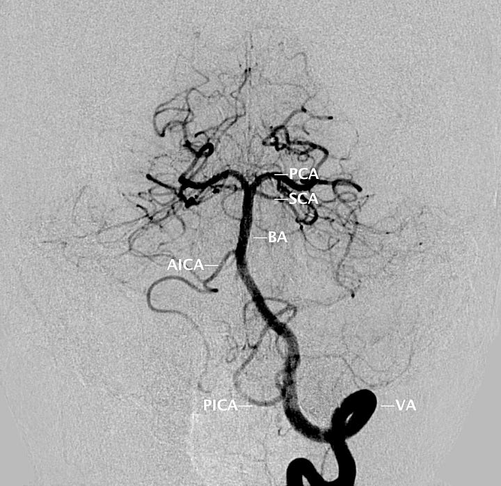
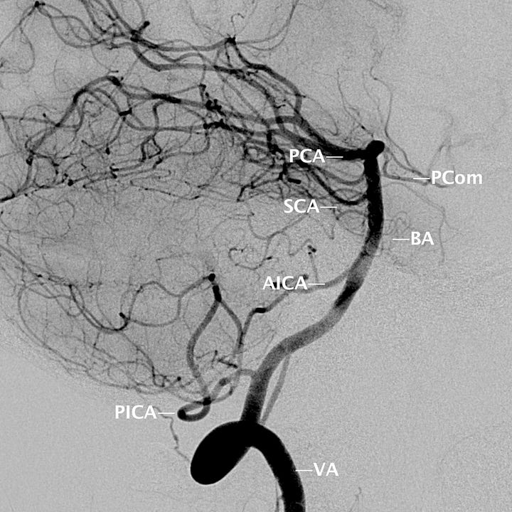
Vertebrobasilar anatomy
VA: Vertebral artery PICA: Posterior inferior cerebellar artery BA: Basilar artery AICA: Anterior inferior cerebellar artery SCA: Superior cerebellar artery PCA: Posterior cerebral artery PCom: Posterior communicating artery.
Note that the third cranal nerve is located adjacent to the basilar artery, in between the superior cerebellar and posterior cerebral arteries. This is why aneurysms that arise from the origin of the posterior communicating artery sometimes cause oculomotor symptoms (ptosis, pupillary dilatation, and inferior and lateral deviation of the eye with consequent diplopia)


Not much for the vessels I've described. The main variants involve hypoplasia of anterior communicating, posterior communicating, or the proximal segments of the posterior cerebral arteries. But there is of course variation, knowledge of which is critical for neuroangiography and neurosurgery. I would refer you to:
Normal Variants of the Cerebral Circulation at Multidetector CT Angiography
Simon J. Dimmick, BPthy, MBBS • Kenneth C. Faulder, MBBS, FRANZCR
RadioGraphics 2009; 29:1027–1043
How much variation in that vascular anatomy, between patients?