Acute Subdural Hematoma
Motor vehicle accident with left hemiparesis and depressed consciousness


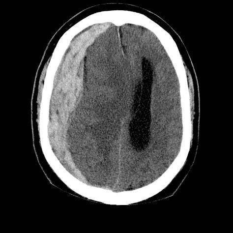
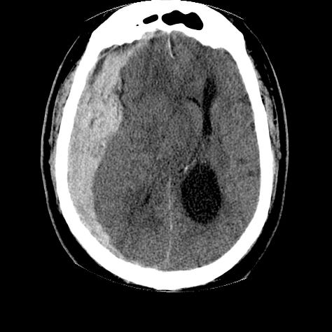
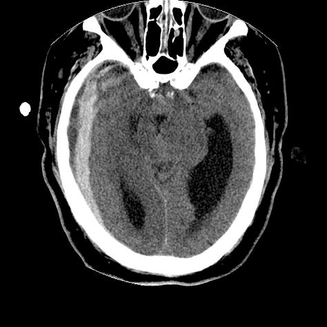
A large right sided blood collection is located between the inner layer of the dura and the arachnoid membrane and extends over the entire cerebral hemisphere on this non-contrast CT. The underlying brain is compressed, with herniation under the falx, herniation through the right tentorial notch, compression of the right lateral ventricle and obstruction (“trapping”) of the left lateral ventricle.
Subdural hematomas are usually due to severe acceleration/deceleration forces and result in shearing of the veins that travel from the brain, across the subdural space to the venous sinuses. They are bounded by the dural reflections: the falx and tentorium. Often associated with significant underlying cerebral injury, prognosis in a subdural hematoma of this size would be poor, even with prompt surgical evacuation.
Acute subdural hematomas are generally hyperdense compared with the brain on CT, and gradually decrease in density as they age. Mixed high and low density hematomas are considered hyper acute and indicate active bleeding.

