Acute cholecystitis occurs when a gallstone or biliary sludge obstructs the cystic duct at the gallbladder neck. Patients present with right upper quadrant pain that radiates to the back, nausea, and anorexia. Early laboratory findings may be normal, but leukocytosis and mild transaminase elevation eventually occur as a result of biliary obstruction and inflammation. Untreated acute cholecystitis can progress to wall ischemia, gangrenous or emphysematous cholecystitis and perforation.
Risk factors include female sex, obesity, pregnancy, age > 40, sickle cell disease, rapid weight loss and use of GLP-1 agonists, particularly at higher doses. Acute or chronic pancreatitis, cholangitis, appendicitis, mesenteric ischemia, cardiac disease renal colic and peptic ulcer disease can mimic clinical symptoms of cholecystitis. Biliary colic refers to right upper quadrant pain that resolves within 6 hours and is due to transient cystic duct obstruction by a gallstone.
Ultrasound is the primary imaging approach in right upper quadrant pain and accurately identifies gallstones and gallbladder inflammation. Gallstones, pericholecystic fluid, luminal distention, gallbladder wall thickening, and pain on compression of the gallbladder under visualization (the sonographic Murphy sign) make up the constellation of supportive findings. The normal gallbladder should be less than 4 cm in diameter, and its wall should be less than 3 mm thick. The best evidence of acute cholecystitis is detection of gallstones in conjunction with a sonographic Murphy sign. Gallbladder wall thickening and pericholecystic fluid are less definitive findings and can be seen in other conditions such as hepatitis, pancreatitis, or adjacent colitis.
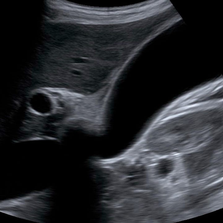
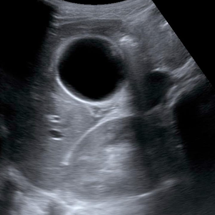
Acute cholecystitis (ultrasound). The gallbladder is markedly distended with minimal wall thickening but without pericholecystic fluid. A shadowing mass (gallstone) is located in the gallbladder neck.
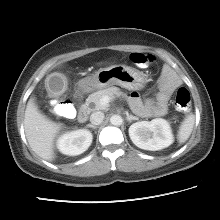
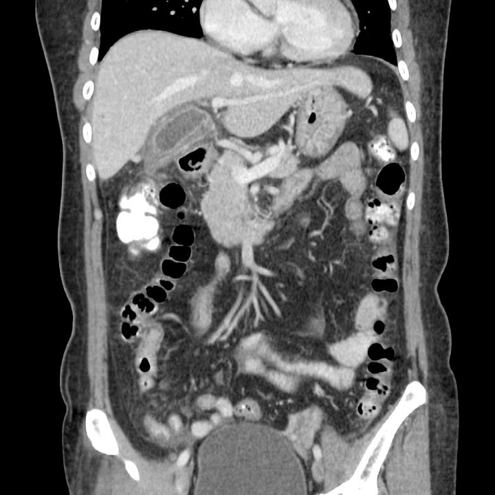
Acute cholecystitis (CT). The gallbladder wall is thickened, with associated pericholecystic inflammatory stranding and pericholecystic fluid. The common bile duct is mildly distended.
Cholecystitis is often diagnosed on CT obtained for generalized abdominal pain or in patients whose ultrasound is non-diagnostic due to obesity or other technical factors. It is less sensitive then ultrasound for detecting gallstones, which may be isodense to bile in some cases. In patients with equivocal ultrasound or CT findings, hepatobiliary iminodiacetic acid (HIDA) scan can be obtained and is considered diagnostic of cholecystitis if the gallbladder does not fill 4 hours after radionuclide administration.
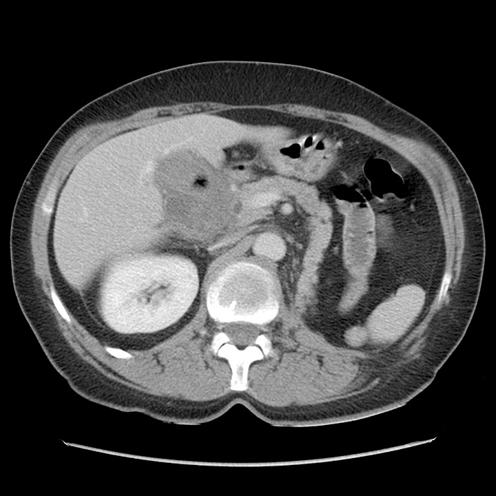
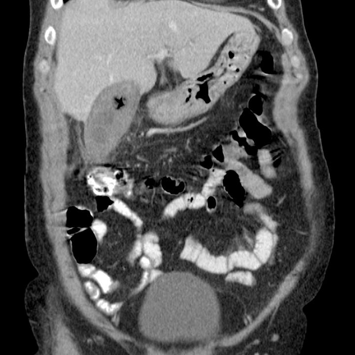
Acute cholecystitis and cholelithiasis. The gallbladder is distended with sludge and near isodense gallstones, one of which contains gas. Mild pericholecystic inflammatory fat stranding.
Management is usually by urgent or early elective laparoscopic cholecystectomy. Gallbladder drainage by percutaneous cholecystostomy can be performed in unstable or high risk patents as a temporizing measure.

